I saw this video over at the Worms & Germs blog. It's a new take on a popular Christmas carol. Enjoy!
Lyrics
On the twelfth day of Christmas my true love gave to me,
Twelve tubs of Purell,
Eleven raccoon roundworms,
Ten cats-a-scratching
Nine hungry hookworms,
Eight dogs-a-biting,
Seven cats with ringworm,
Six big fat dog ticks,
Five cats with fleas.
Four rats with cowpox.
Three tapeworms,
Two toxic turtles,
And a dog with lepto in its pee.
Showing posts with label leptospirosis. Show all posts
Showing posts with label leptospirosis. Show all posts
Monday, December 16, 2013


Tuesday, October 15, 2013
Towards sterilizing immunity against Leptospira with a DNA vaccine
In my previous post, I described the failure of researchers to come up with a conventional protein-based subunit vaccine that confers sterilizing immunity against leptospirosis. What I mean by "conventional" subunit vaccine is a mixture of purified recombinant Leptospira protein with an adjuvant (either aluminum hydroxide or Freund's). Although an antibody response was detected against most proteins tested, immunization failed to prevent kidney colonization in every case, including those animals that survived challenge with lethal strains of Leptosipra. It's becoming clear that the leptospirosis vaccine field must move beyond simple formulations of protein plus adjuvant if sterilizing immunity is desired.
Several labs have explored more modern approaches to delivering leptospirosis vaccines. Odir Dellagostin's group down in Brazil tested the efficacy of DNA vaccines in protecting hamsters against leptospirosis, as described in this paper in Clinical and Vaccine Immunology. Forster and colleagues targeted the Leptospira interrogans LigA and LigB proteins, which are surface proteins that disrupt (or exploit) multiple host functions.
The investigators cloned various fragments of the lengthy ligA and ligB genes downstream of the human cytomegalovirus promoter of the commercial expression plasmid pTARGET. They mixed the plasmid DNA with aluminum hydroxide adjuvant and injected the material into the muscle of the hind leg of hamsters. The animals were given a booster with the same material 21 days later. An IgG immune response was detected against four of the the five Lig protein fragments being tested (see figure below).
21 days after the boost, the animals were challenged with a lethal strain of L. interrogans. The survival curves are shown below. Note that animals immunized with the vector alone (small filled circles) were all dead by day 11.
Among the lig gene fragments, the one expressing the "LigBrep" fragment was effective, protecting five of the eight animals in the group (62.5%) from death. LigBrep comprises amino acid residues 1 through 628 of LigB, whose total length is 1891 residues. Although the survival rate is nothing to get excited over, what distinguishes the LigBrep DNA vaccine from the conventional subunit vaccines tested in earlier studies is that the kidneys from 4 of the 5 survivors were culture negative, indicating that sterilizing immunity was achieved in 80% of the animals that survived infection.
Another notable outcome of the study was that protection was achieved even though the challenge strain and the vaccine's lig gene originated from different Leptospira serovars. One of the problems with killed whole-cell vaccines is that they only protect against Leptospira serovars present in the vaccine formulation because they target LPS, whose structure varies among different serovars. Leptospira proteins tend to be similar in amino acid sequence across different species and are therefore more attractive as vaccines. (The "killed-whole leptospires" control plotted in the graph above was generated from the challenge strain).
So how does DNA vaccination induce sterilizing immunity against Leptospira? As always, more studies are needed to explore this issue, but I will go ahead and speculate. DNA vaccines that are administered by standard injection stimulate a Th1-biased immune response. Studies with cattle have suggested that vaccines must stimulate Th1 immunity to minimize kidney colonization by Leptospira (see this study, for example). Moreover, an earlier study by Dellagostin's group demonstrated sterilizing immunity against L. interrogans in some animals immunized with a Mycobacterium bovis BCG strain that was engineered to express LipL32, the major outer membrane protein of L. interrogans. BCG also stimulates Th1 immunity.
Why would a Th1 response be necessary for sterilizing immunity against Leptospira? Th1 cytokines help steer B cells into producing an IgG isotype that is strongly recognized by Fc receptor on phagocytes. Consequently, bacteria bound by these IgG molecules are engulfed by opsonophagocytosis. During Leptospira infections, opsonophagocytosis clears spirochetes from the circulation during the antibody response, raising the possibility that opsonophagocytosis also leads to sterilizing immunity by vaccines that induce production of the "right" IgG.
Th1 cells are also necessary for cellular immunity, which enhances the killing functions of macrophages so that they can rid themselves of intracellular pathogens. Leptospira is considered to be an extracellular pathogen. Nevertheless, there may be a transient intracellular phase that is critical during infection. Although intracellular Leptospira has not been observed in vivo, L. interrogans is known to survive and replicate in cultured macrophages.
References
Forster KM, Hartwig DD, Seixas FK, Bacelo KL, Amaral M, Hartleben CP, & Dellagostin OA (2013). A conserved region of leptospiral immunoglobulin-like A and B proteins as a DNA vaccine elicits a prophylactic immune response against leptospirosis. Clinical and Vaccine Immunology : CVI, 20 (5), 725-731 PMID: 23486420
Zuerner RL, Alt DP, Palmer MV, Thacker TC, & Olsen SC (2011). A Leptospira borgpetersenii serovar Hardjo vaccine induces a Th1 response, activates NK cells, and reduces renal colonization. Clinical and Vaccine Immunology : CVI, 18 (4), 684-91 PMID: 21288995
Seixas FK, da Silva EF, Hartwig DD, Cerqueira GM, Amaral M, Fagundes MQ, Dossa RG, & Dellagostin OA (2007). Recombinant Mycobacterium bovis BCG expressing the LipL32 antigen of Leptospira interrogans protects hamsters from challenge. Vaccine, 26 (1), 88-95 PMID: 18063449
Toma C, Okura N, Takayama C, & Suzuki T (2011). Characteristic features of intracellular pathogenic Leptospira in infected murine macrophages. Cellular microbiology, 13 (11), 1783-1192 PMID: 21819516
Related posts
Several labs have explored more modern approaches to delivering leptospirosis vaccines. Odir Dellagostin's group down in Brazil tested the efficacy of DNA vaccines in protecting hamsters against leptospirosis, as described in this paper in Clinical and Vaccine Immunology. Forster and colleagues targeted the Leptospira interrogans LigA and LigB proteins, which are surface proteins that disrupt (or exploit) multiple host functions.
The investigators cloned various fragments of the lengthy ligA and ligB genes downstream of the human cytomegalovirus promoter of the commercial expression plasmid pTARGET. They mixed the plasmid DNA with aluminum hydroxide adjuvant and injected the material into the muscle of the hind leg of hamsters. The animals were given a booster with the same material 21 days later. An IgG immune response was detected against four of the the five Lig protein fragments being tested (see figure below).
 |
| Figure 2 from Forster et al., 2013. Sera were drawn before immunization and after the first and second immunizations with pTARGET-based lig plasmid DNA. Purified recombinant protein encoded by each plasmid was used as antigen in ELISAs. Left, middle, and right bar for each DNA: before immunization, after first immunization, and after second immunization, respectively. |
21 days after the boost, the animals were challenged with a lethal strain of L. interrogans. The survival curves are shown below. Note that animals immunized with the vector alone (small filled circles) were all dead by day 11.
 |
| Figure 3 from Forster et al., 2013. |
Among the lig gene fragments, the one expressing the "LigBrep" fragment was effective, protecting five of the eight animals in the group (62.5%) from death. LigBrep comprises amino acid residues 1 through 628 of LigB, whose total length is 1891 residues. Although the survival rate is nothing to get excited over, what distinguishes the LigBrep DNA vaccine from the conventional subunit vaccines tested in earlier studies is that the kidneys from 4 of the 5 survivors were culture negative, indicating that sterilizing immunity was achieved in 80% of the animals that survived infection.
Another notable outcome of the study was that protection was achieved even though the challenge strain and the vaccine's lig gene originated from different Leptospira serovars. One of the problems with killed whole-cell vaccines is that they only protect against Leptospira serovars present in the vaccine formulation because they target LPS, whose structure varies among different serovars. Leptospira proteins tend to be similar in amino acid sequence across different species and are therefore more attractive as vaccines. (The "killed-whole leptospires" control plotted in the graph above was generated from the challenge strain).
So how does DNA vaccination induce sterilizing immunity against Leptospira? As always, more studies are needed to explore this issue, but I will go ahead and speculate. DNA vaccines that are administered by standard injection stimulate a Th1-biased immune response. Studies with cattle have suggested that vaccines must stimulate Th1 immunity to minimize kidney colonization by Leptospira (see this study, for example). Moreover, an earlier study by Dellagostin's group demonstrated sterilizing immunity against L. interrogans in some animals immunized with a Mycobacterium bovis BCG strain that was engineered to express LipL32, the major outer membrane protein of L. interrogans. BCG also stimulates Th1 immunity.
Why would a Th1 response be necessary for sterilizing immunity against Leptospira? Th1 cytokines help steer B cells into producing an IgG isotype that is strongly recognized by Fc receptor on phagocytes. Consequently, bacteria bound by these IgG molecules are engulfed by opsonophagocytosis. During Leptospira infections, opsonophagocytosis clears spirochetes from the circulation during the antibody response, raising the possibility that opsonophagocytosis also leads to sterilizing immunity by vaccines that induce production of the "right" IgG.
Th1 cells are also necessary for cellular immunity, which enhances the killing functions of macrophages so that they can rid themselves of intracellular pathogens. Leptospira is considered to be an extracellular pathogen. Nevertheless, there may be a transient intracellular phase that is critical during infection. Although intracellular Leptospira has not been observed in vivo, L. interrogans is known to survive and replicate in cultured macrophages.
References
Forster KM, Hartwig DD, Seixas FK, Bacelo KL, Amaral M, Hartleben CP, & Dellagostin OA (2013). A conserved region of leptospiral immunoglobulin-like A and B proteins as a DNA vaccine elicits a prophylactic immune response against leptospirosis. Clinical and Vaccine Immunology : CVI, 20 (5), 725-731 PMID: 23486420
Zuerner RL, Alt DP, Palmer MV, Thacker TC, & Olsen SC (2011). A Leptospira borgpetersenii serovar Hardjo vaccine induces a Th1 response, activates NK cells, and reduces renal colonization. Clinical and Vaccine Immunology : CVI, 18 (4), 684-91 PMID: 21288995
Seixas FK, da Silva EF, Hartwig DD, Cerqueira GM, Amaral M, Fagundes MQ, Dossa RG, & Dellagostin OA (2007). Recombinant Mycobacterium bovis BCG expressing the LipL32 antigen of Leptospira interrogans protects hamsters from challenge. Vaccine, 26 (1), 88-95 PMID: 18063449
Toma C, Okura N, Takayama C, & Suzuki T (2011). Characteristic features of intracellular pathogenic Leptospira in infected murine macrophages. Cellular microbiology, 13 (11), 1783-1192 PMID: 21819516
Related posts
- Is sterilizing immunity against Leptospira possible with protein subunit vaccines?
- A new attentuated leptospirosis vaccine protects hamsters from lethal infection by more than one serovar of Leptospira
Sunday, September 15, 2013
Is sterilizing immunity against Leptospira possible with protein subunit vaccines?
With complete bacterial genome sequences now available, "reverse vaccinology" can be conducted to identify proteins that can function as subunit vaccines. The "gene first" approach of reverse vaccinology relies upon computer analysis of the genome sequence to identify encoded proteins with features common to known surface-exposed and secreted bacterial proteins. The selected genes can then be cloned and expressed as recombinant proteins. The proteins, which may number in the hundreds, are then purified for vaccine testing in the animal model appropriate for the bacterial pathogen. Reverse vaccinology has been employed successfully to find protective protein antigens against Neisseria meningitidis serogroup B, Streptococcus pneumoniae, group B Streptococcus, Bacillus anthracis, Porphyromonas gingivalis, and other bacterial pathogens (reviewed in this paper).
This approach sounds straightforward but in practice may not always lead to identification of effective subunit vaccines. An important study from Ben Adler's group down in Monash University illustrates the challenges of finding a subunit vaccine that prevents chronic Leptospira infections. They focused on serovar Hardjo, which causes chronic infections in cattle. They selected 263 Hardjo genes that were predicted to encode surface-exposed, secreted, or lipid-modified proteins. Among these they successfully cloned and expressed 223 genes as 238 protein antigens in E. coli. Some genes were expressed as two or more fragments because of their large size. 210 of the 238 (88%) aggregated into inclusion bodies during expression and had to be kept dissolved in urea during their purification. (Strangely, the urea was not removed by dialysis prior to immunization.) The 238 purified proteins were mixed with an aluminum hydroxide adjuvant and injected into hamsters. 169 of the 238 proteins (71%) generated an antibody response, yet none succeeded in preventing colonization of the kidneys following challenge with a Hardjo strain.
It's been hard enough to find leptospiral proteins that protect hamsters from lethal disease when tested as vaccines (see this article for a review), yet Murray and colleagues sought proteins that protected against Leptospira colonization, a more difficult endeavor that has never been achieved with subunit vaccines. The Hardjo strain they used easily colonizes the kidneys yet fails to produce any signs of disease in hamsters. Although several studies have demonstrated that certain versions of the LigA and LigB proteins, when administered as vaccines, protect hamsters and mice from being killed by lethal strains of Leptospira, survivors are left with infected kidneys. A vaccine that protects against disease or death but not infection may be adequate for humans, who eventually clear the spirochetes from their kidneys even following a natural infection (assuming the disease doesn't kill them). However, vaccinated cattle infected with Hardjo may not be able to clear the spirochetes and will continue to shed infectious Leptospira into the environment, placing the entire herd and the workers handling them at risk of infection. Hardjo infections generally don't cause signs of disease in cattle, but they can cause fetal death and drop in milk production in cows.
The choice of adjuvant and destruction of protective conformational epitopes by urea are possible reasons for failure to find a protective antigen. On the other hand, perhaps a different method for delivery of protein antigens into animals should been considered. Stay tuned.
Featured paper
Murray GL, Lo M, Bulach DM, Srikram A, Seemann T, Quinsey NS, Sermswan RW, Allen A, & Adler B (2013). Evaluation of 238 antigens of Leptospira borgpetersenii serovar Hardjo for protection against kidney colonisation. Vaccine, 31 (3), 495-499 PMID: 23176980
Helpful reviews
Dellagostin, O.A., Grassmann, A.A., Hartwig, D.D., Felix, S.R., da Silva, E.F., & McBride, A.J.A. (November 2011). Recombinant vaccines against leptospirosis. Human Vaccines 7(11):1215-1224. DOI: 10.4161/hv.7.11.17944
Serruto, D., Serino, L., Masignani, V., & Pizza, M. (May 26, 2009). Genome-based approaches to develop vaccines against bacterial pathogens. Vaccine 27(25-26):3245-3250. DOI: 10.1016/j.vaccine.2009.01.072
This approach sounds straightforward but in practice may not always lead to identification of effective subunit vaccines. An important study from Ben Adler's group down in Monash University illustrates the challenges of finding a subunit vaccine that prevents chronic Leptospira infections. They focused on serovar Hardjo, which causes chronic infections in cattle. They selected 263 Hardjo genes that were predicted to encode surface-exposed, secreted, or lipid-modified proteins. Among these they successfully cloned and expressed 223 genes as 238 protein antigens in E. coli. Some genes were expressed as two or more fragments because of their large size. 210 of the 238 (88%) aggregated into inclusion bodies during expression and had to be kept dissolved in urea during their purification. (Strangely, the urea was not removed by dialysis prior to immunization.) The 238 purified proteins were mixed with an aluminum hydroxide adjuvant and injected into hamsters. 169 of the 238 proteins (71%) generated an antibody response, yet none succeeded in preventing colonization of the kidneys following challenge with a Hardjo strain.
It's been hard enough to find leptospiral proteins that protect hamsters from lethal disease when tested as vaccines (see this article for a review), yet Murray and colleagues sought proteins that protected against Leptospira colonization, a more difficult endeavor that has never been achieved with subunit vaccines. The Hardjo strain they used easily colonizes the kidneys yet fails to produce any signs of disease in hamsters. Although several studies have demonstrated that certain versions of the LigA and LigB proteins, when administered as vaccines, protect hamsters and mice from being killed by lethal strains of Leptospira, survivors are left with infected kidneys. A vaccine that protects against disease or death but not infection may be adequate for humans, who eventually clear the spirochetes from their kidneys even following a natural infection (assuming the disease doesn't kill them). However, vaccinated cattle infected with Hardjo may not be able to clear the spirochetes and will continue to shed infectious Leptospira into the environment, placing the entire herd and the workers handling them at risk of infection. Hardjo infections generally don't cause signs of disease in cattle, but they can cause fetal death and drop in milk production in cows.
The choice of adjuvant and destruction of protective conformational epitopes by urea are possible reasons for failure to find a protective antigen. On the other hand, perhaps a different method for delivery of protein antigens into animals should been considered. Stay tuned.
Featured paper
Murray GL, Lo M, Bulach DM, Srikram A, Seemann T, Quinsey NS, Sermswan RW, Allen A, & Adler B (2013). Evaluation of 238 antigens of Leptospira borgpetersenii serovar Hardjo for protection against kidney colonisation. Vaccine, 31 (3), 495-499 PMID: 23176980
Helpful reviews
Dellagostin, O.A., Grassmann, A.A., Hartwig, D.D., Felix, S.R., da Silva, E.F., & McBride, A.J.A. (November 2011). Recombinant vaccines against leptospirosis. Human Vaccines 7(11):1215-1224. DOI: 10.4161/hv.7.11.17944
Serruto, D., Serino, L., Masignani, V., & Pizza, M. (May 26, 2009). Genome-based approaches to develop vaccines against bacterial pathogens. Vaccine 27(25-26):3245-3250. DOI: 10.1016/j.vaccine.2009.01.072
Thursday, January 3, 2013
Spirochete research: 2012 in review
Here are some of my favorite spirochete papers from 2012. Direct access to all research articles (some behind a paywall) is provided via the DOI links. Where present, the links above the citations lead to my blog posts about the studies.
The Leptospira interrogans LigB protein protects the spirochete from complement by capturing complement regulatory proteins.
A B. burgdorferi lipase with hemolytic activity in vitro:
L. interrogans sheath protein homologs that are not needed for flagellar sheath formation:
A critical analysis of a study that demonstrated persistence of B. burgdorferi in infected rhesus monkeys that were treated with antibiotics:
Presenting flawed studies directly to the public to bypass the scientific peer-review process:
PATHOGENESIS
Two distinct regions of the Borrelia burgdorferi BBK32 lipoprotein sequentially mediate binding to the vessel wall in vivo during escape of the spirochete from the bloodstream.- Moriarty TJ, Shi , Lin Y-P, Ebady R, Zhou H, Odisho T, Hardy P-O, Salman-Dilgimen A, Wu J, Weening EH, Skare JT, Kubes P, Leong J, and Chaconas G (December 2012). Vascular binding of a pathogen under shear force through mechanistically distinct sequential interactions with host macromolecules. Molecular Microbiology 86(5):1116-1131. DOI: 10.1111/mmi.12045
The Leptospira interrogans LigB protein protects the spirochete from complement by capturing complement regulatory proteins.
- Castiblanco-Valencia MM, Fraga TR, da Silva LB, Monaris D, Abreu PAE, Strobel S, Jozsi M, Isaac L, and Barbosa AS (March 15, 2012). Leptospiral immunoglobulin-like proteins interact with human complement regulators factor H, FHL-1, FHR-1, and C4BP. The Journal of Infectious Diseases 205(6):995-1004. DOI: 10.1093/infdis/jir875
- Choy HA (July 2012). Multiple activities of LigB potentiate virulence of Leptospira interrogans: inhibition of alternative and classical pathways of complement. PLoS One 7(7):e41566. DOI: 10.1371/journal.pone.0041566
A B. burgdorferi lipase with hemolytic activity in vitro:
- Shaw DK, Hyde JA, and Skare JT (January 2012). The BB0646 protein demonstrates lipase and haemolytic activity associated with Borrelia burgdorferi, the aetiological agent of Lyme disease. Molecular Microbiology 83(2):319-334. DOI: 10.1111/j.1365-2958.2011.07932.x
TRANSMISSION
Borrelia burgdorferi needs the alternative sigma factor RpoS to flee from the tick's midgut- Dunham-Ems SM, Caimano MJ, Eggers CH, and Radolf JD (February 2012). Borrelia burgdorferi requires the alternative sigma factor RpoS for Dissemination within the vector during tick-to-mammal transmission. PLoS Pathogens 8(2):e1002532. DOI: 10.1371/journal.ppat.1002532
MOTILITY
Video microscopy of B. burgdorferi swimming around in gelatin and mouse tissue:- Harman MW, Dunham-Ems SM, Caimano MJ, Belperron AA, Bockenstedt LK, Fu HC, Radolf JD, and Wolgemuth CW (February 21, 2012). The heterogeneous motility of the Lyme disease spirochete in gelatin mimics dissemination through tissue. Proceedings of the National Academy of Sciences USA 109(8):3059-3064. DOI: 10.1073/pnas.1114362109
L. interrogans sheath protein homologs that are not needed for flagellar sheath formation:
- Lambert A, Picardeau M, Haake DA, Sermswan RW, Srikram A, Adler B, and Murray GA (June 2012). FlaA proteins in Leptospira interrogans are essential for motility and virulence but are not required for formation of the flagellum sheath. Infection and Immunity 80(6):2019-2025. DOI: 10.1128/IAI.00131-12
ULTRASTRUCTURE
A close look at the ultrastructure of Leptospira without the artifacts generated by conventional electron microscopy:- Raddi G, Morado DR, Yan J, Haake DA, Yang XF, and Liu J (March 2012). Three-dimensional structures of pathogenic and saprophytic Leptospira species revealed by cryo-electron tomography. Journal of Bacteriology 194(6):1299-1306. DOI: 10.1128/JB.06474-11
METAL TOXICITY
B. burgdorferi BicA, a protein that protects the spirochete from the toxic effects of copper and iron:- Wang P, Lutton A, Olesik J, Vali H, and Li X (December 2012). A novel iron- and copper-binding protein in the Lyme disease spirochaete. Molecular Microbiology 86(6):1441-1451. DOI: 10.1111/mmi.12068
ANTIBIOTIC THERAPY
Inflammatory spirochete debris left behind following antibiotic treatment for Lyme disease- Bockenstedt LK, Gonzalez DG, Haberman AM, and Belperron AA (July 2, 2012). Spirochete antigens persist near cartilage after murine Lyme borreliosis therapy. The Journal of Clinical Investigation 122(7):2652-2660. DOI: 10.1172/JCI58813
A critical analysis of a study that demonstrated persistence of B. burgdorferi in infected rhesus monkeys that were treated with antibiotics:
- Wormser GP, Baker PJ, O'Connell S, Pachner AR, Schwartz I, and Shapiro ED (July 2012). Critical analysis of treatment trials of rhesus macaques infected with Borrelia burgdorferi reveals important flaws in experimental design. Vector-borne and Zoonotic Diseases 12(7):535-538. DOI: 10.1089/vbz.2012.1012
- Embers ME, Barthold SW, Borda JT, Bowers L, Doyle L, Hodzic E, Jacobs MB, Hasenkampf NR, Martin DS, Narasimhan S, Phillippi-Falkenstein KM, Purcell JE, Ratterree MS, and Philipp MT (January 2012). Persistence of Borrelia burgdorferi in rhesus macaques following antibiotic treatment of disseminated infection. PLoS One 7(1):e29914. DOI: 10.1371/journal.pone.0029914
- Wormser GP, Daniels TJ, Bittker S, Cooper D, Wang G, and Pavia CS (March 15, 2012). Failure of topical antibiotics to prevent disseminated Borrelia burgdorferi infection following a tick bite in C3H/HeJ mice. The Journal of Infectious Diseases 205(6):991-994. DOI: 10.1093/infdis/jir382
DIAGNOSTICS
Not so golden? Microscopic agglutination test for diagnosis of leptospirosis- Limmathurotsakul D, Turner EL, Wuthiekanun V, Thaipadungpanit J, Suputtamongkol Y, Chierakul W, Smythe LD, Day NPJ, Cooper B, and Peacock SJ (August 1, 2012). Fool’s gold: Why imperfect reference tests are undermining the evaluation of novel diagnostics: A reevaluation of 5 diagnostic tests for leptospirosis. Clinical Infectious Diseases 55(3):322-331. DOI: 10.1093/cid/cis403
ECOLOGY
Do nonspiral spirochetes help clean our environment?- Caro-Quintero A, Ritalahti KM, Cusick KD, Loffler FE, and Konstandtinidis KT (May/June 2012). The chimeric genome of Sphaerochaeta: Nonspiral spirochetes that break with the prevalent dogma in spirochete biology. mBio 3(3):e00025-12. DOI: 10.1128/mBio.00025-12
- Ritalahti KM, Justicia-Leon SD, Cusick KD, Ramos-Hernandez N, Rubin M, Dornbush J, and Loffler FE (January 2012). Sphaerochaeta globosa gen. nov., sp. nov. and Sphaerochaeta pleomorpha sp. nov., free-living, spherical spirochetes. International Journal of Systematic and Evolutionary Microbiology 62(Pt 1):210-216. DOI: 10.1099/ijs.0.023986-0
BIOFILM
Biofilms of the Lyme disease spirochete- Sapi E, Bastian SL, Mpoy CM, Scott S, Rattelle A, Pabbati N, Poruri A, Burugu D, Theophilus PAS, Pham TV, Data A, Dhaliwal NK, MacDonald A, Rossi MJ, Sinha SK, and Luecke DF (October 2012). Characterization of biofilm formation by Borrelia burgdorferi in vitro. PLoS One 7(10):e48277. DOI: 10.1371/journal.pone.0048277
HISTORY
Looking for the syphilis spirochete in ancient bones- Montiel R, Solorzano E, Diaz N, Alvarez-Sandoval BA, Gonzalez-Ruiz M, Canadas MP, Simoes N, Isidro A, and Malgosa A (May 2012). Neonate human remains: A window of opportunity to the molecular analysis of syphilis. PLoS One 7(5):e36371. DOI: 10.1371/journal.pone.0036371
Presenting flawed studies directly to the public to bypass the scientific peer-review process:
- Armelagos GJ, Zuckerman MK, and Harper KN (March 2012). The science behind pre-Columbian evidence of syphilis in Europe: research by documentary. Evolutionary Anthropology 21(2):50-57. DOI: 10.1002/evan.20340
REVIEWS
Here are two excellent review articles that appeared during the past year:- Radolf JD, Caimano MJ, Stevenson B, and Hu LT (February 2012). Of ticks, mice and men: understanding the dual-host lifestyle of Lyme disease spirochaetes. Nature Reviews Microbiology 10(2):87-99. DOI: 10.1038/nrmicro2714
- Charon NW, Cockburn A, Li C, Liu J, Miller KA, Miller MR, Motaleb MA, and Wolgemuth CW (2012). The unique paradigm of spirochete motility and chemotaxis. Annual Reviews of Microbiology 66:349-370. DOI: 10.1146/annurev-micro-092611-150145
Monday, November 26, 2012
A post-Thanksgiving story of leptospirosis
I'm about half way through 1491, a book that gives readers a view of the Americas before Columbus showed up. It also describes the devastating impact that foreign infectious diseases had on the native population as Europeans explored the New World.
One chapter tells the story of Tisquantum (Squanto), who lived in the village of Patuxet, one of the many Indian communities thriving along the coast of New England at the time. In 1614 Thomas Hunt, a British slave trader, kidnapped Tisquantum and other Indians and shipped them to Spain. Fortunately, Tisquantum was rescued by Spanish priests before he could be sold. After convincing the priests to let him return home, he left for London, where he learned English while staying at a shipbuilder's home, and eventually made his way back to North America. As he sailed down the New England shoreline in 1619 on a British ship, he realized that the world familiar to him had vanished. A mysterious disease had wiped out 90% of the population of coastal New England. When he arrived at his home village of Patuxet, he found it deserted. Tisquantum was soon captured and sent to Massasoit, the leader of the Wampanoag confederacy, which encompassed Patuxet. Massasoit did not trust Tisquantum because of his recent association with the British, yet he would later use him as a translator in a negotiation that turned out to be a pivotal event in American history.

The epidemic had been blamed at one time or another on smallpox, the plague, yellow fever, typhus, and hepatitis. As I've mentioned before, a recent analysis has added leptospirosis to the list of suspects. The symptoms and signs of leptospirosis match those reported from first-hand accounts of the mystery ailment. Here's a post on the Slate website about the epidemic. I'm glad to see that the story is getting attention from popular news sites.
Leptospirosis can be deadly, but could it account for the devastating 90% motality rate of the 1616-1619 epidemic? A hypervirulent strain of Leptospira or genetic susceptibility of the Indians could be an explanation. However, the authors of the study thought that the most critical factor was the Indian lifestyle, which brought them into repeated contact with Leptospira in the environment. The Europeans who fished nearby were spared because they did not engage in activities that exposed them to Leptospira. Therefore, only the Indians contracted the illness, according to the hypothesis.
Whatever its cause, it's hard to overstate the significance of the epidemic. Prior to 1616, the New England native communities traded with the Europeans and even welcomed them for brief stays. However, all attempts by the foreigners to establish permanent settlements were fiercely resisted. Coastal New England was well defended by the large native population. The Wampanoag confederacy became especially hostile towards the Europeans after having their citizens abducted.
By the time the Mayflower landed in Patuxet (Plymouth) in December of 1620, the thinking of the Wampanoag had changed. Their depleted population was vulnerable to attack by their longtime enemies to the west, the Narragansett, who remained untouched by the epidemic. To forestall an attack, Massasoit felt that the best course of action was to form an alliance with the Pilgrims rather than expel them. In the spring of 1621, with Tisquantum serving as the translator, Massasoit arranged a peace treaty with the Pilgrims.
Reference
Marr J.S. & Cathey J.T. (2010). New hypothesis for cause of epidemic among Native Americans, New England, 1616–1619, Emerging Infectious Diseases, 16 (2) 281-286. DOI: 10.3201/eid1602.090276
Related post
One chapter tells the story of Tisquantum (Squanto), who lived in the village of Patuxet, one of the many Indian communities thriving along the coast of New England at the time. In 1614 Thomas Hunt, a British slave trader, kidnapped Tisquantum and other Indians and shipped them to Spain. Fortunately, Tisquantum was rescued by Spanish priests before he could be sold. After convincing the priests to let him return home, he left for London, where he learned English while staying at a shipbuilder's home, and eventually made his way back to North America. As he sailed down the New England shoreline in 1619 on a British ship, he realized that the world familiar to him had vanished. A mysterious disease had wiped out 90% of the population of coastal New England. When he arrived at his home village of Patuxet, he found it deserted. Tisquantum was soon captured and sent to Massasoit, the leader of the Wampanoag confederacy, which encompassed Patuxet. Massasoit did not trust Tisquantum because of his recent association with the British, yet he would later use him as a translator in a negotiation that turned out to be a pivotal event in American history.
The epidemic had been blamed at one time or another on smallpox, the plague, yellow fever, typhus, and hepatitis. As I've mentioned before, a recent analysis has added leptospirosis to the list of suspects. The symptoms and signs of leptospirosis match those reported from first-hand accounts of the mystery ailment. Here's a post on the Slate website about the epidemic. I'm glad to see that the story is getting attention from popular news sites.
Leptospirosis can be deadly, but could it account for the devastating 90% motality rate of the 1616-1619 epidemic? A hypervirulent strain of Leptospira or genetic susceptibility of the Indians could be an explanation. However, the authors of the study thought that the most critical factor was the Indian lifestyle, which brought them into repeated contact with Leptospira in the environment. The Europeans who fished nearby were spared because they did not engage in activities that exposed them to Leptospira. Therefore, only the Indians contracted the illness, according to the hypothesis.
 |
| Figure 3 from Marr and Cathey, 2010. |
Whatever its cause, it's hard to overstate the significance of the epidemic. Prior to 1616, the New England native communities traded with the Europeans and even welcomed them for brief stays. However, all attempts by the foreigners to establish permanent settlements were fiercely resisted. Coastal New England was well defended by the large native population. The Wampanoag confederacy became especially hostile towards the Europeans after having their citizens abducted.
By the time the Mayflower landed in Patuxet (Plymouth) in December of 1620, the thinking of the Wampanoag had changed. Their depleted population was vulnerable to attack by their longtime enemies to the west, the Narragansett, who remained untouched by the epidemic. To forestall an attack, Massasoit felt that the best course of action was to form an alliance with the Pilgrims rather than expel them. In the spring of 1621, with Tisquantum serving as the translator, Massasoit arranged a peace treaty with the Pilgrims.
Reference
Marr J.S. & Cathey J.T. (2010). New hypothesis for cause of epidemic among Native Americans, New England, 1616–1619, Emerging Infectious Diseases, 16 (2) 281-286. DOI: 10.3201/eid1602.090276
Related post
Sunday, July 22, 2012
Not so golden? Microscopic agglutination test for diagnosis of leptospirosis
The microscopic agglutination test (MAT) is designated the "gold standard" for the laboratory diagnosis of leptospirosis, a spirochete disease that can cause severe illness if not promptly treated. Although imperfect, MAT is used as the benchmark when the performance of another diagnostic test for leptospirosis is being assessed. It is also used to determine the prevalence of leptospirosis in a population. How imperfect is MAT? A recent study by Limmathurotsakul and colleagues, published in the journal Clinical Infectious Diseases, claims that its performance is much worse than scientists previously thought.
MAT involves mixing serial dilutions of patient sera with live suspensions of Leptospira. If agglutinating antibodies against Leptospira are present, the spirochetes will clump. The clumps can be seen by darkfield microscopy. Although the idea behind MAT is simple to understand, the technique itself is cumbersome. Since agglutinating antibodies react best with the specific Leptospira serovar infecting the patient, cultures of at least one serovar from each of the ~20 major Leptospira serogroups must be maintained. To perform the assay, each serum dilution is mixed individually with a suspension from each culture and examined by microscopy one at a time. The assay is time consuming, laborious, and potentially hazardous to laboratory personnel. For these reasons MAT is not routinely employed for diagnostic testing outside of the research setting.
The performance of a diagnostic test is judged by its sensitivity and specificity. The problem with leptospirosis is figuring out how many actually have the disease so that the sensitivity can be calculated accurately. Since the sensitivity of culture is poor, researchers rely on antibody tests such as MAT to identify leptospirosis cases. This approach assumes that the sensitivity and specificity of MAT are 100%.
In general there are two problems with using antibody tests for diagnostics. The first is that it takes time for the immune system to generate enough antibody that can be detected. The second problem is that those with previous exposure to the pathogen will test positive even if they are not currently infected. To minimize these problems, patients with the signs and symptoms of leptospirosis are deemed to have a positive MAT if they fulfill one of the following criteria.
Since some false negative MAT cases can be identified by culture, one way to calculate the sensitivity of MAT is to add the number of MAT-positive and culture-positive (but MAT-negative) cases together to estimate the number of patients with leptospirosis and then calculate the percentage of MAT-positive cases among these patients. Limmathurotsakul and colleagues performed these calculations with data from their four earlier studies conducted in Thailand. A total of 413 patients tested positive by MAT or culture (or both). They found that the sensitivity of MAT was 86%-96% across the four sets of data. The remaining 4-14% were false negatives, having tested positive by culture but not by MAT.
The authors next calculated the true sensitivity and specificity of MAT with a statistical tool called latent class analysis, which does not assume any perfect gold standard. Since there is no perfect test, the true disease status of each patient is the unknown or "latent" variable. Results from multiple diagnostic tests are related to the latent variable using statistical models. The calculations go beyond the scope of this blog post, but the bottom line is that the true sensitivity and specificity of each diagnostic test can be estimated with these models. In addition to MAT and culture, the authors tested some of their patients with an immunofluorescence assay (IFA), lateral flow test (LF), and/or PCR. Latent class analysis is more powerful when the diagnostic tests being evaluated detect different features of the infection. MAT, IFA, and LF are antibody tests, and culture and PCR detect the pathogen itself.
The sensitivity of MAT calculated by this method turned out to be only 49.8%, much lower than the 86%-96% calculated using the standard method that assumes a perfect gold standard. The sensitivity of culture alone was 10.5%. Combining culture with MAT did not help much; the sensitivity of the combined approach was only 55.5%. The low sensitivity of "MAT plus culture" suggests that the specificities calculated for the alternative tests may be underestimated by the standard method. This is because some of the many false-negative cases may be correctly identified as having leptospirosis by the alternative tests. This turned out to be the case for two of the tests. Specificities for all tests were over 95% by latent class analysis. However, the specificities for PCR (82.5%) and the lateral flow test (70.5%) were lower when "MAT plus culture" was assumed to be the perfect gold standard.
You can see that the accuracy of alternative leptospirosis tests is underestimated when MAT (or MAT plus culture) is assumed to be the perfect reference test. Another implication of the study is that the prevalence of leptospirosis has been underestimated, at least in Thailand. The only other study to evaluate the performance of MAT by latent class analysis was conducted by the CDC here in the U.S almost a decade ago. In contrast to the Limmathurostsakul study, the CDC study determined that the sensitivity of MAT was high, at 98.2%. There were many differences between the two studies, including the patient population, the alternative tests evaluated, the time interval between collection of paired sera, and the number of serovars included for MAT. The poor performance of MAT in the Thailand study may therefore not be a universal finding.
References
Limmathurotsakul D, Turner EL, Wuthiekanun V, Thaipadungpanit J, Suputtamongkol Y, Chierakul W, Smythe LD, Day NPJ, Cooper B, Peacock SJ (August 2012). Fool's gold: why imperfect reference tests are undermining the evaluation of novel diagnostics: a reevaluation of 5 diagnostic tests for leptospirosis. Clinical Infectious Diseases 55(3):322-331. DOI: 10.1093/cid/cis403
Bajani MD, Ashford DA, Bragg SL, Woods CW, Aye T, Spiegel RA, Plikaytis BD, Perkins BA, Phelan M, Levett PN, and Weyant RS (February 2003). Evaluation of four commercially available rapid serologic tests for diagnosis of leptospirosis. Journal of Clinical Microbiology 41(2):803-809. DOI: 10.1128/JCM.41.2.803-809.2003
MAT involves mixing serial dilutions of patient sera with live suspensions of Leptospira. If agglutinating antibodies against Leptospira are present, the spirochetes will clump. The clumps can be seen by darkfield microscopy. Although the idea behind MAT is simple to understand, the technique itself is cumbersome. Since agglutinating antibodies react best with the specific Leptospira serovar infecting the patient, cultures of at least one serovar from each of the ~20 major Leptospira serogroups must be maintained. To perform the assay, each serum dilution is mixed individually with a suspension from each culture and examined by microscopy one at a time. The assay is time consuming, laborious, and potentially hazardous to laboratory personnel. For these reasons MAT is not routinely employed for diagnostic testing outside of the research setting.
The performance of a diagnostic test is judged by its sensitivity and specificity. The problem with leptospirosis is figuring out how many actually have the disease so that the sensitivity can be calculated accurately. Since the sensitivity of culture is poor, researchers rely on antibody tests such as MAT to identify leptospirosis cases. This approach assumes that the sensitivity and specificity of MAT are 100%.
In general there are two problems with using antibody tests for diagnostics. The first is that it takes time for the immune system to generate enough antibody that can be detected. The second problem is that those with previous exposure to the pathogen will test positive even if they are not currently infected. To minimize these problems, patients with the signs and symptoms of leptospirosis are deemed to have a positive MAT if they fulfill one of the following criteria.
- At least a four-fold increase in MAT titer between paired sera.
- At least a 1:400 MAT titer when only a single specimen is available. This cutoff is sometimes adjusted based on the prevalence of leptospirosis in the population being examined.
Since some false negative MAT cases can be identified by culture, one way to calculate the sensitivity of MAT is to add the number of MAT-positive and culture-positive (but MAT-negative) cases together to estimate the number of patients with leptospirosis and then calculate the percentage of MAT-positive cases among these patients. Limmathurotsakul and colleagues performed these calculations with data from their four earlier studies conducted in Thailand. A total of 413 patients tested positive by MAT or culture (or both). They found that the sensitivity of MAT was 86%-96% across the four sets of data. The remaining 4-14% were false negatives, having tested positive by culture but not by MAT.
The authors next calculated the true sensitivity and specificity of MAT with a statistical tool called latent class analysis, which does not assume any perfect gold standard. Since there is no perfect test, the true disease status of each patient is the unknown or "latent" variable. Results from multiple diagnostic tests are related to the latent variable using statistical models. The calculations go beyond the scope of this blog post, but the bottom line is that the true sensitivity and specificity of each diagnostic test can be estimated with these models. In addition to MAT and culture, the authors tested some of their patients with an immunofluorescence assay (IFA), lateral flow test (LF), and/or PCR. Latent class analysis is more powerful when the diagnostic tests being evaluated detect different features of the infection. MAT, IFA, and LF are antibody tests, and culture and PCR detect the pathogen itself.
The sensitivity of MAT calculated by this method turned out to be only 49.8%, much lower than the 86%-96% calculated using the standard method that assumes a perfect gold standard. The sensitivity of culture alone was 10.5%. Combining culture with MAT did not help much; the sensitivity of the combined approach was only 55.5%. The low sensitivity of "MAT plus culture" suggests that the specificities calculated for the alternative tests may be underestimated by the standard method. This is because some of the many false-negative cases may be correctly identified as having leptospirosis by the alternative tests. This turned out to be the case for two of the tests. Specificities for all tests were over 95% by latent class analysis. However, the specificities for PCR (82.5%) and the lateral flow test (70.5%) were lower when "MAT plus culture" was assumed to be the perfect gold standard.
You can see that the accuracy of alternative leptospirosis tests is underestimated when MAT (or MAT plus culture) is assumed to be the perfect reference test. Another implication of the study is that the prevalence of leptospirosis has been underestimated, at least in Thailand. The only other study to evaluate the performance of MAT by latent class analysis was conducted by the CDC here in the U.S almost a decade ago. In contrast to the Limmathurostsakul study, the CDC study determined that the sensitivity of MAT was high, at 98.2%. There were many differences between the two studies, including the patient population, the alternative tests evaluated, the time interval between collection of paired sera, and the number of serovars included for MAT. The poor performance of MAT in the Thailand study may therefore not be a universal finding.
References
Limmathurotsakul D, Turner EL, Wuthiekanun V, Thaipadungpanit J, Suputtamongkol Y, Chierakul W, Smythe LD, Day NPJ, Cooper B, Peacock SJ (August 2012). Fool's gold: why imperfect reference tests are undermining the evaluation of novel diagnostics: a reevaluation of 5 diagnostic tests for leptospirosis. Clinical Infectious Diseases 55(3):322-331. DOI: 10.1093/cid/cis403
Bajani MD, Ashford DA, Bragg SL, Woods CW, Aye T, Spiegel RA, Plikaytis BD, Perkins BA, Phelan M, Levett PN, and Weyant RS (February 2003). Evaluation of four commercially available rapid serologic tests for diagnosis of leptospirosis. Journal of Clinical Microbiology 41(2):803-809. DOI: 10.1128/JCM.41.2.803-809.2003
Saturday, March 31, 2012
qPCR testing for Leptospira infection reveals sex differences in bacterial load
The pitfall of serological testing is that patients test negative during the early days of infection because time is needed for the immune response to ramp up antibody production against the pathogen. Unfortunately clinical laboratories are stuck with serological methods for detecting Leptospira since direct tests for the spirochete are not available.
Since Leptospira has to enter the bloodstream to spread throughout the body, PCR testing of blood is one tool that may aid prompt diagnosis of leptospirosis. In a study published in Clinical Infectious Diseases, Agampodi and colleagues conducted quantitative real-time PCR (qPCR) on patient sera collected during a 2008 leptospirosis outbreak in Sri Lanka, where leptospirosis is endemic. With a reported annual incidence of 5.4 cases per 100,000, Sri Lanka has the sixth highest incidence of leptospirosis in the world.
The acute-phase sera tested for the study came from patients with confirmed leptospirosis. The patients had clinical evidence of leptospirosis, and a second blood sample drawn at least a week later indicated that they had rising antibody titers or had seroconverted.
The PCR primers targeted the DNA encoding the 16S rRNA of Leptospira. The sensitivity of qPCR turned out to be 51% (25/49) with acute-phase sera collected during the first ten days of illness. This doesn't sound impressive at all, but the sensitivity of the microscopic agglutination test (MAT) was much worse. MAT testing is done by mixing dilutions of serum with suspensions of Leptospira. If anti-Leptospira antibodies are present, they will cause the bacteria to clump. The sensitivity of MAT with acute-phase sera collected up to 15 days after symptoms began was a miserable 18% (13/73). Past studies conducted in Sri Lanka and elsewhere around the world have also demonstrated poor performance of serological testing for leptospirosis. Although the specificity of qPCR wasn't determined, it's clear that qPCR is an improvement over serological testing for the prompt diagnosis of leptospirosis.
One interesting observation came out of the qPCR data. Men who were qPCR positive had a higher bacterial load than women who tested positive (median of 15,640 bacteria/ml in men vs. 5,611 bacteria/ml in women, P = 0.022, Mann-Whitney U test). Based on this observation, the authors raised the possibility that men are biologically more susceptible to leptospirosis than women. On the other hand, the difference in bacterial load may have nothing to do with biology. It may simply reflect differences in when men and women sought medical care, or it may mean men were exposed to environmental sources that were more heavily contaminated with Leptospira.
A 2007 study conducted in Germany revealed sex differences in leptospirosis severity. Men diagnosed with leptospirosis were more likely than women to have hemorrhage, jaundice, and renal impairment, all signs of severe disease (see table below). The difference in disease severity could not be accounted for by differences in exposure risk, the infecting serogroup, or interaction with the health care system. Could the difference be due to biological variation between the sexes?
Note: Here's a recent post in the blog Camp Other that examined sex differences in another disease of spirochetes, Lyme disease.
Main references
Agampodi SB, Matthias MA, Moreno AC, and Vinetz JM (March 12, 2012). Utility of quantitative polymerase chain reaction in leptospirosis diagnosis: association of level of leptospiremia and clinical manifestations in Sri Lanka. Clinical Infectious Diseases (published online ahead of print February 21, 2012). DOI: 10.1093/cid/cis035
Jansen A, Stark K, Schneider T, and Schöneberg I (May 1, 2007). Sex differences in clinical leptospirosis in Germany: 1997-2005. Clinical Infectious Diseases 44(9):e69-e72. DOI: 10.1016/j.ijid.2007.09.011
Other references
Pappas G, Papadimitriou P, Siozopoulou V, Christou L, and Akritidis N (July 2008). The globalization of leptospirosis: worldwide incidence trend. International Journal of Infectious Diseases 12(4):351-357. DOI: 10.1016/j.ijid.2007.09.011
Reller ME, Bodinayake C, Nagahawatte A, Devasiri V, Kodikara-Arachichi W, Strouse JJ, Flom JE, Dumier JS, and Woods CW (September 9, 2011). Leptospirosis as frequent cause of acute febrile illness in southern Sri Lanka. Emerging Infectious Diseases 17(9):1678-1684. DOI: 10.3201/eid1709.100915
Since Leptospira has to enter the bloodstream to spread throughout the body, PCR testing of blood is one tool that may aid prompt diagnosis of leptospirosis. In a study published in Clinical Infectious Diseases, Agampodi and colleagues conducted quantitative real-time PCR (qPCR) on patient sera collected during a 2008 leptospirosis outbreak in Sri Lanka, where leptospirosis is endemic. With a reported annual incidence of 5.4 cases per 100,000, Sri Lanka has the sixth highest incidence of leptospirosis in the world.
The acute-phase sera tested for the study came from patients with confirmed leptospirosis. The patients had clinical evidence of leptospirosis, and a second blood sample drawn at least a week later indicated that they had rising antibody titers or had seroconverted.
The PCR primers targeted the DNA encoding the 16S rRNA of Leptospira. The sensitivity of qPCR turned out to be 51% (25/49) with acute-phase sera collected during the first ten days of illness. This doesn't sound impressive at all, but the sensitivity of the microscopic agglutination test (MAT) was much worse. MAT testing is done by mixing dilutions of serum with suspensions of Leptospira. If anti-Leptospira antibodies are present, they will cause the bacteria to clump. The sensitivity of MAT with acute-phase sera collected up to 15 days after symptoms began was a miserable 18% (13/73). Past studies conducted in Sri Lanka and elsewhere around the world have also demonstrated poor performance of serological testing for leptospirosis. Although the specificity of qPCR wasn't determined, it's clear that qPCR is an improvement over serological testing for the prompt diagnosis of leptospirosis.
One interesting observation came out of the qPCR data. Men who were qPCR positive had a higher bacterial load than women who tested positive (median of 15,640 bacteria/ml in men vs. 5,611 bacteria/ml in women, P = 0.022, Mann-Whitney U test). Based on this observation, the authors raised the possibility that men are biologically more susceptible to leptospirosis than women. On the other hand, the difference in bacterial load may have nothing to do with biology. It may simply reflect differences in when men and women sought medical care, or it may mean men were exposed to environmental sources that were more heavily contaminated with Leptospira.
A 2007 study conducted in Germany revealed sex differences in leptospirosis severity. Men diagnosed with leptospirosis were more likely than women to have hemorrhage, jaundice, and renal impairment, all signs of severe disease (see table below). The difference in disease severity could not be accounted for by differences in exposure risk, the infecting serogroup, or interaction with the health care system. Could the difference be due to biological variation between the sexes?
 |
| Table 1 from Jansen et al., 2007. OR adjusted for age. |
Note: Here's a recent post in the blog Camp Other that examined sex differences in another disease of spirochetes, Lyme disease.
Main references
Agampodi SB, Matthias MA, Moreno AC, and Vinetz JM (March 12, 2012). Utility of quantitative polymerase chain reaction in leptospirosis diagnosis: association of level of leptospiremia and clinical manifestations in Sri Lanka. Clinical Infectious Diseases (published online ahead of print February 21, 2012). DOI: 10.1093/cid/cis035
Jansen A, Stark K, Schneider T, and Schöneberg I (May 1, 2007). Sex differences in clinical leptospirosis in Germany: 1997-2005. Clinical Infectious Diseases 44(9):e69-e72. DOI: 10.1016/j.ijid.2007.09.011
Other references
Pappas G, Papadimitriou P, Siozopoulou V, Christou L, and Akritidis N (July 2008). The globalization of leptospirosis: worldwide incidence trend. International Journal of Infectious Diseases 12(4):351-357. DOI: 10.1016/j.ijid.2007.09.011
Reller ME, Bodinayake C, Nagahawatte A, Devasiri V, Kodikara-Arachichi W, Strouse JJ, Flom JE, Dumier JS, and Woods CW (September 9, 2011). Leptospirosis as frequent cause of acute febrile illness in southern Sri Lanka. Emerging Infectious Diseases 17(9):1678-1684. DOI: 10.3201/eid1709.100915
Sunday, October 23, 2011
Life after leptospirosis, a pilot study
The signs and symptoms associated with acute human leptospirosis are well known. Less is known about how well patients manage after they begin recovering from the illness. Most who survive the illness, even those who had severe illness, appear to regain full health eventually. However careful examination may reveal subtle deficiencies in organ function.2 Moreover the spectrum of health problems that could crop up later is not known. The only late complication of leptospirosis that has been well documented is uveitis, which can lead to eye pain and vision problems.3 Old case studies have also reported recurring headaches, general malaise, and even dementia, aggression, depression, and psychosis in those surviving leptospirosis,4,5 although these observations would need to be confirmed in well-designed clinical studies.
To gain a better understanding of the long-term outcomes of leptospirosis, a large systematic study is needed to monitor the health of leptospirosis patients as they heal from the illness. Such a study was attempted by Spichler and colleagues.1 Their pilot study targeted patients hospitalized with leptospirosis at any time from December 2008 to May 2009 in São Paulo, Brazil, where leptospirosis is endemic. There were 180 patients hospitalized with leptospirosis during this period, but for a variety of reasons only 47 of the 180 could be enrolled in the study. All 47 had been treated for at least seven days with antibiotics (penicillin or ceftriaxone) during their hospitalization, and all had their diagnosis confirmed by serologic testing. 17 of the 47 had severe leptospirosis, defined as those who had experienced jaundice, kidney failure, bleeding (hemorrhage), pulmonary (lung) involvement, or shock while hospitalized. Fortunately no patient died.
The first outpatient visit was conducted an average of 22 ± 12 days after the patients were discharged from the hospital. 23 of the 47 (49%) continued to experience one or more of the following health problems: general malaise, headache, muscle pain, dizziness, bronchitis, and abdominal pain. Three patients still had jaundice.
Only 22 of the 47 patients came back for a second visit, which took place a mean of 40 ± 21 days after they were discharged from the hospital. Two of the 22 patients continued to suffer from medical problems.
One individual was experiencing general malaise, which wasn't a problem for him before being stricken with leptospirosis. He did always have high blood pressure, which may or may not have been a contributing factor to his malaise. An ECG showed some abnormalities with his heartbeat. Leptospira is known to cause myocarditis, an inflammation of the heart muscle. This patient continued to suffer from profound general malaise when examined one year later.
The second patient started to experience panic attacks between the two clinic visits. Were the panic attacks related to his earlier bout with leptospirosis? It's difficult to conclude anything from this single patient. Although the patient had severe leptospirosis, did not suffer from any of the known neurologic features of leptospirosis during his acute illness6 and had not been diagnosed with any neurologic or psychiatric condition before contracting leptospirosis. Panic disorder has never been described in leptospirosis patients in the scientific literature.
Since this was a pilot study, the investigators probably lacked the resources to address the problems that cropped up during the study. As pointed out by the authors, the major limitations of the study were that few of the eligible patients were enrolled in the study and that many dropped out between visits, resulting in a potentially biased sampling of leptospirosis patients. Additionally the follow-up visits did not include any laboratory testing to detect lingering functional deficiencies in the kidney, liver, and other organs. For these reason it's impossible to make any definitive conclusions about the recovery of these subjects. Despite the preliminary nature of this pilot study, the possible outcomes of acute leptospirosis identified in this study and earlier case studies beg for a future prospective study with a larger number of individuals living in an area where leptospirosis is endemic. A longer time frame for follow up is also necessary since uveitis can first appear up to four years after recovery from leptospirosis.3
Any future study should also follow those with mild or asymptomatic Leptospira infections. The reason is that the long-term outcome of mild disease is unknown. Since those with mild or asymptomatic infections are unlikely to seek medical attention, identifying such individuals will require investigators to monitor healthy high-risk individuals by serologic testing so that the newly infected could be identified as those with increasing anti-Leptospira antibody titers.
In light of the recent discovery of chronically infected individuals in the Peruvian Amazon,7 it would also be prudent to test the urine of healthy individuals for Leptospira being shed from the kidney tubules so that the findings of the Peruvian study could be confirmed. The long-term effects of chronic infection, if any, could also be identified.
Where could a future study examining the long-term outcomes of Leptospira infection be conducted? Brazil and India may be best suited for this type of study. Leptospirosis is highly endemic in those countries, and multiple research teams are already investigating the epidemiology of leptospirosis in those countries.
10-30-2011, edits for clarity.
Featured paper
1. Spichler A., Athanazio D, Seguro AC, and Vinetz JM (July 2011). Outpatient follow-up of patients hospitalized for acute leptospirosis. International Journal of Infectious Diseases 15(7):e486-e490. DOI: 10.1016/j.ijid.2011.03.020
References
2. de Francesco Daher E, Zanetta DMT, and Abdulkader RCRM (September 2004). Pattern of renal function recovery after leptospirosis acute renal failure. Nephron, Clinical Practice 98(1):c8-c14. DOI: 10.1159/000079922
3. Shukla D, Rathinam SR, and Cunningham ET (Spring 2010). Leptospiral uveitis in the developing world. International Ophthalmology Clinics 50(2):113-124. DOI:
4. Shpilberg O, Shaked Y, Maier MK, Samra D, and Samra Y (April 1990). Long-term follow-up after leptospirosis. Southern Medical Journal 83(4):405-407. Link
5. Avery TL (July 27, 1983). Leptospirosis and mental illness. New Zealand Medical Journal 96(736):589 (Letter).
6. Panicker JN, Mammachan R, and Jayakumar RV (September 2001). Primary neuroleptospirosis. Postgraduate Medical Journal 77(911):589-590. DOI: 10.1136/pmj.77.911.589
7. Ganoza CA, Matthias MA, Collins-Richards D, Brouwer KC, Cunningham CB, Segura ER, Gilman RH, Gotuzzo E, and Vinetz JM (2006). Determining risk for severe leptospirosis by molecular analysis of environmental surface waters for pathogenic Leptospira. PLoS Medicine 3(8):e308. DOI: 10.1371/journal.pmed.0030308
Related post
To gain a better understanding of the long-term outcomes of leptospirosis, a large systematic study is needed to monitor the health of leptospirosis patients as they heal from the illness. Such a study was attempted by Spichler and colleagues.1 Their pilot study targeted patients hospitalized with leptospirosis at any time from December 2008 to May 2009 in São Paulo, Brazil, where leptospirosis is endemic. There were 180 patients hospitalized with leptospirosis during this period, but for a variety of reasons only 47 of the 180 could be enrolled in the study. All 47 had been treated for at least seven days with antibiotics (penicillin or ceftriaxone) during their hospitalization, and all had their diagnosis confirmed by serologic testing. 17 of the 47 had severe leptospirosis, defined as those who had experienced jaundice, kidney failure, bleeding (hemorrhage), pulmonary (lung) involvement, or shock while hospitalized. Fortunately no patient died.
The first outpatient visit was conducted an average of 22 ± 12 days after the patients were discharged from the hospital. 23 of the 47 (49%) continued to experience one or more of the following health problems: general malaise, headache, muscle pain, dizziness, bronchitis, and abdominal pain. Three patients still had jaundice.
Only 22 of the 47 patients came back for a second visit, which took place a mean of 40 ± 21 days after they were discharged from the hospital. Two of the 22 patients continued to suffer from medical problems.
One individual was experiencing general malaise, which wasn't a problem for him before being stricken with leptospirosis. He did always have high blood pressure, which may or may not have been a contributing factor to his malaise. An ECG showed some abnormalities with his heartbeat. Leptospira is known to cause myocarditis, an inflammation of the heart muscle. This patient continued to suffer from profound general malaise when examined one year later.
The second patient started to experience panic attacks between the two clinic visits. Were the panic attacks related to his earlier bout with leptospirosis? It's difficult to conclude anything from this single patient. Although the patient had severe leptospirosis, did not suffer from any of the known neurologic features of leptospirosis during his acute illness6 and had not been diagnosed with any neurologic or psychiatric condition before contracting leptospirosis. Panic disorder has never been described in leptospirosis patients in the scientific literature.
Since this was a pilot study, the investigators probably lacked the resources to address the problems that cropped up during the study. As pointed out by the authors, the major limitations of the study were that few of the eligible patients were enrolled in the study and that many dropped out between visits, resulting in a potentially biased sampling of leptospirosis patients. Additionally the follow-up visits did not include any laboratory testing to detect lingering functional deficiencies in the kidney, liver, and other organs. For these reason it's impossible to make any definitive conclusions about the recovery of these subjects. Despite the preliminary nature of this pilot study, the possible outcomes of acute leptospirosis identified in this study and earlier case studies beg for a future prospective study with a larger number of individuals living in an area where leptospirosis is endemic. A longer time frame for follow up is also necessary since uveitis can first appear up to four years after recovery from leptospirosis.3
Any future study should also follow those with mild or asymptomatic Leptospira infections. The reason is that the long-term outcome of mild disease is unknown. Since those with mild or asymptomatic infections are unlikely to seek medical attention, identifying such individuals will require investigators to monitor healthy high-risk individuals by serologic testing so that the newly infected could be identified as those with increasing anti-Leptospira antibody titers.
In light of the recent discovery of chronically infected individuals in the Peruvian Amazon,7 it would also be prudent to test the urine of healthy individuals for Leptospira being shed from the kidney tubules so that the findings of the Peruvian study could be confirmed. The long-term effects of chronic infection, if any, could also be identified.
Where could a future study examining the long-term outcomes of Leptospira infection be conducted? Brazil and India may be best suited for this type of study. Leptospirosis is highly endemic in those countries, and multiple research teams are already investigating the epidemiology of leptospirosis in those countries.
10-30-2011, edits for clarity.
Featured paper
1. Spichler A., Athanazio D, Seguro AC, and Vinetz JM (July 2011). Outpatient follow-up of patients hospitalized for acute leptospirosis. International Journal of Infectious Diseases 15(7):e486-e490. DOI: 10.1016/j.ijid.2011.03.020
References
2. de Francesco Daher E, Zanetta DMT, and Abdulkader RCRM (September 2004). Pattern of renal function recovery after leptospirosis acute renal failure. Nephron, Clinical Practice 98(1):c8-c14. DOI: 10.1159/000079922
3. Shukla D, Rathinam SR, and Cunningham ET (Spring 2010). Leptospiral uveitis in the developing world. International Ophthalmology Clinics 50(2):113-124. DOI:
4. Shpilberg O, Shaked Y, Maier MK, Samra D, and Samra Y (April 1990). Long-term follow-up after leptospirosis. Southern Medical Journal 83(4):405-407. Link
5. Avery TL (July 27, 1983). Leptospirosis and mental illness. New Zealand Medical Journal 96(736):589 (Letter).
6. Panicker JN, Mammachan R, and Jayakumar RV (September 2001). Primary neuroleptospirosis. Postgraduate Medical Journal 77(911):589-590. DOI: 10.1136/pmj.77.911.589
7. Ganoza CA, Matthias MA, Collins-Richards D, Brouwer KC, Cunningham CB, Segura ER, Gilman RH, Gotuzzo E, and Vinetz JM (2006). Determining risk for severe leptospirosis by molecular analysis of environmental surface waters for pathogenic Leptospira. PLoS Medicine 3(8):e308. DOI: 10.1371/journal.pmed.0030308
Related post
Sunday, May 1, 2011
A new attenuated leptospirosis vaccine protects hamsters from lethal infection by more than one serovar of Leptospira
Scientists have demonstrated that a new attenuated leptospirosis vaccine protects laboratory hamsters from being killed by Leptospira, even when the challenge and vaccine strains belong to different serovars (immune types).1,2 This is the first leptospirosis vaccine to confer complete cross-protection against lethal infection by a serovar different from the one used for immunization.
The leptospirosis vaccines that are out on the market are still formulated with killed Leptospira or sometimes their outer membrane. These traditional vaccines are administered primarily to dogs, cattle, and pigs. Human leptospirosis vaccines are not available in most countries, even in areas where leptospirosis is endemic.
New types of leptospirosis vaccines are needed since the traditional killed vaccines are flawed. One problem is that immunity is serovar specific. For this reason a vaccine must contain all the serovars that the target population may encounter. Even when the vaccine manufacturers figure out which serovars are circulating, a new serovar may emerge, rendering the vaccine ineffective as the new serovar spreads through the susceptible population. The vaccine must then be reformulated at substantial cost.
This is exactly what happened to the leptospirosis vaccines that are given to dogs.3 The early canine vaccines, first available in the 1970s, contained the serovars Canicola and Icterohemorrhagiae. These vaccines worked fine until the late 1980s or so, when new serovars started to appear in infected dogs, even in those that had been vaccinated. Since then vaccine makers have added the serovars Grippotyphosa and Pomona to their vaccines. Nevertheless with over 200 pathogenic serovars of Leptospira lurking out there, we don't know when or which additional serovars will emerge in the future.
It would be nice to have a single leptospirosis vaccine formulation that would protect against all serovars. The protective effect of traditional vaccines is due to antibodies generated against lipopolysaccharide (LPS), whose structure differs among the serovars of Leptospira. Since immunization elicits antibodies that recognize the LPS of only the serovars included in the vaccine, vaccinated individuals remain susceptible to infection by other serovars.
To get around this problem, scientists have been testing individual Leptospira surface proteins as potential vaccines in rodent models of leptospirosis. Leptospira surface proteins tend to be antigenically conserved among the different serovars: antibodies generated against a protein from one serovar often reacts against the same protein expressed by other serovars. According to many studies the LipL32 and Lig surface lipoproteins, when delivered as recombinant proteins, naked DNA, or by microbial vectors (adenovirus and Mycobacterium bovis), apparently protected hamsters or guinea pigs from lethal infection by Leptospira. Unfortunately one of the leaders in the leptospirosis field, Ben Adler (also an author of the two featured papers), has questioned the interpretation of these studies.4 He points out that the challenge strains used in several studies were not sufficiently lethal, making it easier to observe a protective effect of the vaccine. Moreover some studies claimed statistically significant protective effects of the protein vaccine when in fact there was none upon Adler's reanalysis of the data. The only protein to convincingly exert a protective effect in an appropriate animal model was LigA5 although the ability of the LigA subunit vaccine to cross-protect against different serovars of Leptospira has yet to be tested. However there is one major problem with using LigA as a vaccine--not all Leptospira strains have the ligA gene.6
In the two studies described here the investigators took a step back from looking at individual proteins and developed an attenuated strain to use for immunization. The properties of the attenuated strain, designated M1352, are described in the paper authored by Murray and colleagues.2 The M1352 strain was not developed by the classic approach of continuously growing and passaging the bacteria in culture until they lost their ability to cause disease. Instead the strain was one of a large collection of mutants generated by random transposon mutagenesis of L. interrogans serovar Manilae. The M1352 strain had the transposon inserted in a gene located in a large cluster of genes encoding enzymes that assemble LPS. The mutation had subtle effects on the reactivity of M1352 with various antibodies raised against leptospiral LPS, suggesting that the LPS structure itself was somehow changed in M1352 when compared with the wild-type Manilae strain.
Since LPS is a crucial surface component that interacts with the host, it was not too surprising that M1352 was not able to cause lethal infections like its wild-type Manilae parent. When they infected hamsters with the M1352 strain, the spirochetes were unable to kill the hamsters or even establish an infection in the kidneys. Despite the efficient clearance of M1352, the Leptospira lingered long enough in the hamsters to provoke an antibody response. Western blots of L. interrogans lysates revealed strong reactivity of antibodies from the M1352-infected hamsters to a number of proteins. Because the M1352 strain generated an antibody response without establishing an infection, the authors decided to test the weakened strain as a vaccine in the hamster model in a follow-up study.1
In the second study, Srikram and colleagues1 demonstrated that immunization of hamsters with a single dose of live M1352 was more effective than a dose of heat-killed wild-type strain in protecting hamsters from being killed by the wild-type Manilae strain. The M1352 vaccine also did a better job in preventing colonization of the kidneys by the spirochete and in minimizing lung hemorrhage than the heat-killed vaccine.
When they challenged the vaccinated hamsters with a different serovar, a Pomona strain, all the hamsters immunized with live M1352 survived whereas 60% of animals immunized with heat-killed wild-type Manilae perished. However the M1352 vaccine didn't work perfectly. Although all hamsters immunized with live M1352 survived the challenge with the Pomona strain, the kidneys from 90% of the animals were culture positive, and 90% had hemorrhaged lungs. Nevertheless this is the first time that complete protection from death was observed following challenge of vaccinated animals by a serovar unrelated to the vaccine strain. They also showed that the M1352 vaccine had to be administered alive. Heat-killed or chemically-killed M1352 vaccine failed to protect hamsters from lethal infection.
The investigators next tried to figure out which component of the M1352 strain was the protective cross-reactive antigen targeted by the hamster's immune system. They wondered whether the live M1352 and heat-killed wild-type Manilae vaccines generated antibody responses to different proteins. When they probed separate two-dimensional blots of L. interrogans membrane preparations of serovar Pomona with antibodies from hamsters immunized with M1352 and heat-killed wild-type Manilae, a number of protein spots lit up. Most proteins, including LipL32, reacted with both sets of antibodies. These proteins are unlikely to account for the cross-protection conferred by the M1352 vaccine since the presence of these antibodies in the hamsters immunized with heat-killed Manilae failed to protect the animals from being killed by the Pomona strain. On the other hand, four Pomona proteins were recognized only by hamsters receiving the attenuated vaccine:
Featured papers
1. Srikram A, Zhang K, Bartpho T, Lo M, Hoke DE, Sermswan RW, Adler B, and Murray GL (March 15, 2011). Cross-protective immunity against leptospriosis elicited by a live, attenuated lipopolysaccharide mutant. Journal of Infectious Diseases 203(6):870-879. DOI: 10.1093/infdis/jiq12
2. Murray GL, Amporn S, Henry R, Hartskeerl RA, Sermswan RW, and Adler B (November 2010). Mutations affecting Leptospira interrogans lipopolysaccharide attenuate virulence. Molecular Microbiology 78(3): 701-709. DOI: 10.1111/j.1365-2958.2010.07360.x
Helpful references
3. Guerra MA (February 15, 2009). Leptospirosis. Journal of the American Veterinary Medical Association 234(4):472-478. DOI: 10.2460/javma.234.4.472
4. Adler B and de la Pena Moctezuma (January 27, 2010). Leptospira and leptospirosis. Veterinary Microbiology 140(3-4):287-296. DOI: 10.1016/j.vetmic.2009.03.012
5. Silva, ÉF, Medeiros MA, McBride AJA, Matsunaga J, Esteves GS, Ramos JGR, Santos CS, Croda J, Homma A, Dellagostin OA, Haake DA, Reis MG, and Ko AI (August 14, 2007). The terminal portion of leptospiral immunoglobulin-like protein LigA confers protective immunity against lethal infection in the hamster model of leptospirosis. Vaccine 25(33):6277-6286. DOI: 10.1016/j.vaccine.2007.05.053
6. McBride AJA, Cerqueira GM, Suchard MA, Moreira MA, Zuerner RL, Reis MG, Haake DA, Ko AI, and Dellagostin OA (March 2009). Infection, Genetics and Evolution 9(2):196-205. DOI: 10.1016/j.meegid.2008.10.012
7. Ristow P, Bourhy P, da Cruz McBride FW, Figueira CP, Huerre M, Ave P, Girons IS, Ko AI, and Picardeau M (July 2007). The OmpA-like protein Loa22 is essential for leptospiral virulence. PLoS Pathogens3(7):e97. DOI: 10.1371/journal.ppat.0030097
8.Pinne M and Haake DA (June 2009). A comprehensive approach to identification of surface-exposed, outer membrane-spanning proteins of Leptospira interrogans. PLoS One 4(6):e6071. DOI: 10.1371/journal.pone.0006071
The leptospirosis vaccines that are out on the market are still formulated with killed Leptospira or sometimes their outer membrane. These traditional vaccines are administered primarily to dogs, cattle, and pigs. Human leptospirosis vaccines are not available in most countries, even in areas where leptospirosis is endemic.
New types of leptospirosis vaccines are needed since the traditional killed vaccines are flawed. One problem is that immunity is serovar specific. For this reason a vaccine must contain all the serovars that the target population may encounter. Even when the vaccine manufacturers figure out which serovars are circulating, a new serovar may emerge, rendering the vaccine ineffective as the new serovar spreads through the susceptible population. The vaccine must then be reformulated at substantial cost.
This is exactly what happened to the leptospirosis vaccines that are given to dogs.3 The early canine vaccines, first available in the 1970s, contained the serovars Canicola and Icterohemorrhagiae. These vaccines worked fine until the late 1980s or so, when new serovars started to appear in infected dogs, even in those that had been vaccinated. Since then vaccine makers have added the serovars Grippotyphosa and Pomona to their vaccines. Nevertheless with over 200 pathogenic serovars of Leptospira lurking out there, we don't know when or which additional serovars will emerge in the future.
It would be nice to have a single leptospirosis vaccine formulation that would protect against all serovars. The protective effect of traditional vaccines is due to antibodies generated against lipopolysaccharide (LPS), whose structure differs among the serovars of Leptospira. Since immunization elicits antibodies that recognize the LPS of only the serovars included in the vaccine, vaccinated individuals remain susceptible to infection by other serovars.
To get around this problem, scientists have been testing individual Leptospira surface proteins as potential vaccines in rodent models of leptospirosis. Leptospira surface proteins tend to be antigenically conserved among the different serovars: antibodies generated against a protein from one serovar often reacts against the same protein expressed by other serovars. According to many studies the LipL32 and Lig surface lipoproteins, when delivered as recombinant proteins, naked DNA, or by microbial vectors (adenovirus and Mycobacterium bovis), apparently protected hamsters or guinea pigs from lethal infection by Leptospira. Unfortunately one of the leaders in the leptospirosis field, Ben Adler (also an author of the two featured papers), has questioned the interpretation of these studies.4 He points out that the challenge strains used in several studies were not sufficiently lethal, making it easier to observe a protective effect of the vaccine. Moreover some studies claimed statistically significant protective effects of the protein vaccine when in fact there was none upon Adler's reanalysis of the data. The only protein to convincingly exert a protective effect in an appropriate animal model was LigA5 although the ability of the LigA subunit vaccine to cross-protect against different serovars of Leptospira has yet to be tested. However there is one major problem with using LigA as a vaccine--not all Leptospira strains have the ligA gene.6
In the two studies described here the investigators took a step back from looking at individual proteins and developed an attenuated strain to use for immunization. The properties of the attenuated strain, designated M1352, are described in the paper authored by Murray and colleagues.2 The M1352 strain was not developed by the classic approach of continuously growing and passaging the bacteria in culture until they lost their ability to cause disease. Instead the strain was one of a large collection of mutants generated by random transposon mutagenesis of L. interrogans serovar Manilae. The M1352 strain had the transposon inserted in a gene located in a large cluster of genes encoding enzymes that assemble LPS. The mutation had subtle effects on the reactivity of M1352 with various antibodies raised against leptospiral LPS, suggesting that the LPS structure itself was somehow changed in M1352 when compared with the wild-type Manilae strain.
Since LPS is a crucial surface component that interacts with the host, it was not too surprising that M1352 was not able to cause lethal infections like its wild-type Manilae parent. When they infected hamsters with the M1352 strain, the spirochetes were unable to kill the hamsters or even establish an infection in the kidneys. Despite the efficient clearance of M1352, the Leptospira lingered long enough in the hamsters to provoke an antibody response. Western blots of L. interrogans lysates revealed strong reactivity of antibodies from the M1352-infected hamsters to a number of proteins. Because the M1352 strain generated an antibody response without establishing an infection, the authors decided to test the weakened strain as a vaccine in the hamster model in a follow-up study.1
In the second study, Srikram and colleagues1 demonstrated that immunization of hamsters with a single dose of live M1352 was more effective than a dose of heat-killed wild-type strain in protecting hamsters from being killed by the wild-type Manilae strain. The M1352 vaccine also did a better job in preventing colonization of the kidneys by the spirochete and in minimizing lung hemorrhage than the heat-killed vaccine.
When they challenged the vaccinated hamsters with a different serovar, a Pomona strain, all the hamsters immunized with live M1352 survived whereas 60% of animals immunized with heat-killed wild-type Manilae perished. However the M1352 vaccine didn't work perfectly. Although all hamsters immunized with live M1352 survived the challenge with the Pomona strain, the kidneys from 90% of the animals were culture positive, and 90% had hemorrhaged lungs. Nevertheless this is the first time that complete protection from death was observed following challenge of vaccinated animals by a serovar unrelated to the vaccine strain. They also showed that the M1352 vaccine had to be administered alive. Heat-killed or chemically-killed M1352 vaccine failed to protect hamsters from lethal infection.
The investigators next tried to figure out which component of the M1352 strain was the protective cross-reactive antigen targeted by the hamster's immune system. They wondered whether the live M1352 and heat-killed wild-type Manilae vaccines generated antibody responses to different proteins. When they probed separate two-dimensional blots of L. interrogans membrane preparations of serovar Pomona with antibodies from hamsters immunized with M1352 and heat-killed wild-type Manilae, a number of protein spots lit up. Most proteins, including LipL32, reacted with both sets of antibodies. These proteins are unlikely to account for the cross-protection conferred by the M1352 vaccine since the presence of these antibodies in the hamsters immunized with heat-killed Manilae failed to protect the animals from being killed by the Pomona strain. On the other hand, four Pomona proteins were recognized only by hamsters receiving the attenuated vaccine:
- Loa22, the only surface protein known to be essential for L. interrogans to cause lethal infections7
- a homolog of GspG, a component of the type II secretion system
- LA1939, a possible lipoprotein of unknown function
- OmpL36, a surface-exposed outer membrane protein of unknown function8
Featured papers
1. Srikram A, Zhang K, Bartpho T, Lo M, Hoke DE, Sermswan RW, Adler B, and Murray GL (March 15, 2011). Cross-protective immunity against leptospriosis elicited by a live, attenuated lipopolysaccharide mutant. Journal of Infectious Diseases 203(6):870-879. DOI: 10.1093/infdis/jiq12
2. Murray GL, Amporn S, Henry R, Hartskeerl RA, Sermswan RW, and Adler B (November 2010). Mutations affecting Leptospira interrogans lipopolysaccharide attenuate virulence. Molecular Microbiology 78(3): 701-709. DOI: 10.1111/j.1365-2958.2010.07360.x
Helpful references
3. Guerra MA (February 15, 2009). Leptospirosis. Journal of the American Veterinary Medical Association 234(4):472-478. DOI: 10.2460/javma.234.4.472
4. Adler B and de la Pena Moctezuma (January 27, 2010). Leptospira and leptospirosis. Veterinary Microbiology 140(3-4):287-296. DOI: 10.1016/j.vetmic.2009.03.012
5. Silva, ÉF, Medeiros MA, McBride AJA, Matsunaga J, Esteves GS, Ramos JGR, Santos CS, Croda J, Homma A, Dellagostin OA, Haake DA, Reis MG, and Ko AI (August 14, 2007). The terminal portion of leptospiral immunoglobulin-like protein LigA confers protective immunity against lethal infection in the hamster model of leptospirosis. Vaccine 25(33):6277-6286. DOI: 10.1016/j.vaccine.2007.05.053
6. McBride AJA, Cerqueira GM, Suchard MA, Moreira MA, Zuerner RL, Reis MG, Haake DA, Ko AI, and Dellagostin OA (March 2009). Infection, Genetics and Evolution 9(2):196-205. DOI: 10.1016/j.meegid.2008.10.012
7. Ristow P, Bourhy P, da Cruz McBride FW, Figueira CP, Huerre M, Ave P, Girons IS, Ko AI, and Picardeau M (July 2007). The OmpA-like protein Loa22 is essential for leptospiral virulence. PLoS Pathogens3(7):e97. DOI: 10.1371/journal.ppat.0030097
8.Pinne M and Haake DA (June 2009). A comprehensive approach to identification of surface-exposed, outer membrane-spanning proteins of Leptospira interrogans. PLoS One 4(6):e6071. DOI: 10.1371/journal.pone.0006071
Sunday, February 20, 2011
Another reason not to own a pet rat
A pair of European doctors wrote about the trials of a 37-year-old man who came down with Weil's disease, a severe form of leptospirosis. The case report appeared in the recent issue of The Lancet Infectious Diseases.
The individual showed up in the emergency room complaining of a sudden fever, muscle pain, and a severe headache. Notable were the signs of jaundice and conjunctival suffusion (reddening of the eye), which suggested leptospirosis.
Shortly after he was admitted, the patient had to be moved to the intensive care unit because his kidneys were failing. Blood tests for Leptospira antibodies came back negative, most likely because it was too early in the infection for antibodies to appear.
An infectious diseases specialist noted that the patient suffered from the classic signs of Weil's disease: kidney failure, jaundice, and an enlarged spleen. Accordingly, the patient was started on intravenous penicillin, the favored treatment for severe leptospirosis. Further blood tests conducted during the second week of treatment finally revealed antibodies to Leptospira. The patient completely healed within several weeks. The only question that remained is how the patient got infected with Leptospira.
At some point the patient admitted that he owned four pet rats. Since rats can silently carry Leptospira in their kidneys, his pets were sacrificed to check their kidneys for the presence of the spirochete. All four kidneys turned out to be culture positive for Leptospira interrogans, serovar Icterohaemorrhagiae or Copenhageni (the typing test couldn't distinguish the two serovars). The patient's antibodies reacted against the same serovars, suggesting that the source of the infection was tainted urine from his pet rats.
Although cases of leptospirosis acquired from pet rats appear to be rare, anyone who wishes to own a rat should be aware that rats can be carriers of a potentially deadly spirochete. This is yet another reason why rats should be obtained from a responsible breeder.
For another case, check out this post from the Worms & Germs Blog (make sure you read the comments for the complete story).
Reference
Jansen, A. and Schneider, T. (February 2011). Weil's disease in a rat owner. The Lancet Infectious Diseases 11(2):152. DOI: 10.1016/S1473-3099(10)70106-7
The individual showed up in the emergency room complaining of a sudden fever, muscle pain, and a severe headache. Notable were the signs of jaundice and conjunctival suffusion (reddening of the eye), which suggested leptospirosis.
 |
| Figure from Jansen and Schneider, 2011 |
An infectious diseases specialist noted that the patient suffered from the classic signs of Weil's disease: kidney failure, jaundice, and an enlarged spleen. Accordingly, the patient was started on intravenous penicillin, the favored treatment for severe leptospirosis. Further blood tests conducted during the second week of treatment finally revealed antibodies to Leptospira. The patient completely healed within several weeks. The only question that remained is how the patient got infected with Leptospira.
At some point the patient admitted that he owned four pet rats. Since rats can silently carry Leptospira in their kidneys, his pets were sacrificed to check their kidneys for the presence of the spirochete. All four kidneys turned out to be culture positive for Leptospira interrogans, serovar Icterohaemorrhagiae or Copenhageni (the typing test couldn't distinguish the two serovars). The patient's antibodies reacted against the same serovars, suggesting that the source of the infection was tainted urine from his pet rats.
Although cases of leptospirosis acquired from pet rats appear to be rare, anyone who wishes to own a rat should be aware that rats can be carriers of a potentially deadly spirochete. This is yet another reason why rats should be obtained from a responsible breeder.
For another case, check out this post from the Worms & Germs Blog (make sure you read the comments for the complete story).
Reference
Jansen, A. and Schneider, T. (February 2011). Weil's disease in a rat owner. The Lancet Infectious Diseases 11(2):152. DOI: 10.1016/S1473-3099(10)70106-7
Sunday, August 1, 2010
The major outer membrane protein of Leptospira interrogans: Not essential for infection?
Because leptospirosis is a potentially fatal disease, it would be worthwhile to figure out which of the many genes on the two chromosomes of Leptospira express products that are essential for infection.
The lipoprotein LipL32 is the most abundant outer membrane protein found in the outer membrane of pathogenic species of Leptosipra. It's been assumed that LipL32 plays an important role in infections for the following reasons:
The survival curves show that the L. interrogans lipL32 mutant was just as lethal to hamsters as the parent L. interrogans with its lipL32 gene intact, irrespective of the infection route. Hence, lipL32 is not necessary for lethal infections of hamsters, at least under the conditions used in this study.
Rats are the natural reservoir hosts of L. interrogans. They can carry the spirochete for years in their kidney tubules without showing any signs of illness. LipL32 could have a role in chronic infections. The investigators therefore tested the ability of the lipL32 mutant to establish a chronic infection in laboratory rats. They found that the lipL32 mutant (M933) was able to colonize the rat kidneys as well as the control M777 strain. Kidneys from all 8 rats inoculated with the lipL32 mutant were culture positive.
At first glance it's surprising that lipL32 was not required for acute or chronic infection. The authors pointed out that the function of LipL32 could be copied by other proteins found on the surface of Leptospira. However, the study could have been strengthened by making two changes. First, since the authors were trying to determine whether lipL32 was necessary for chronic infection, the rats should have been allowed to live for at least a few months before their kidneys were cultured. Instead, the infection was allowed to proceed for only 15 days before the rats were sacrificed. Second, they should have measured the bacterial load in the rat kidneys, either by plating serial dilutions of the kidney homogenates for colonies (although I don't know if this is feasible for Leptospira) or by quantitative PCR. Clearly, more work needs to be done before anyone can conclude that LipL32 is not essential for chronic infection.
Featured paper
Murray G.L., Srikram, A., Hoke, D.E., Wunder Jr., E.A., Henry, R., Lo, M., Zhang, K., Sermswan, R.W., Ko, A.I., and Adler, B. (March 2009). Major surface protein LipL32 is not required for either acute or chronic infection with Leptospira interrogans. Infection and Immunity 77(3):952-958. DOI: 10.1128/IAI.01370-08
Related posts
The lipoprotein LipL32 is the most abundant outer membrane protein found in the outer membrane of pathogenic species of Leptosipra. It's been assumed that LipL32 plays an important role in infections for the following reasons:
- LipL32 is found only in pathogenic species of Leptospira. Nonpathogenic species such as L. biflexa lack the gene encoding LipL32.
- LipL32 peaks out on the surface of Leptospira, where it is available to interact directly with host molecules.
- LipL32 binds (at least weakly) to several components of the extracellular matrix.
- Leptospirosis patients generate a strong antibody response against LipL32.
- The protein sequence of LipL32 among different species of Leptospira is almost identical.
- A lot of metabolic energy must be expended to make the large amounts of LipL32 found in the spirochete.
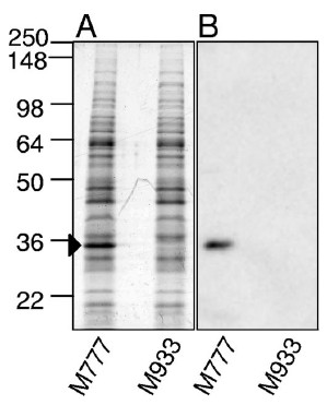 |
| Whole-cell lysates of the L. interrogans lipL32 mutant (M933) and a control strain with the transposon in an intergenic region (M777) were run into SDS-acylamide gels and stained (panel A) or analyzed by Western blotting with LipL32 antiserum (panel B). The M777 strain was demonstrated in an earlier study to be lethal for hamsters. (Figure 1 from Murray et al., 2009.) |
The survival curves show that the L. interrogans lipL32 mutant was just as lethal to hamsters as the parent L. interrogans with its lipL32 gene intact, irrespective of the infection route. Hence, lipL32 is not necessary for lethal infections of hamsters, at least under the conditions used in this study.
 | |||
| Panel A: Groups of 8 hamsters were inoculated with 1,000 leptospires into the abdominal cavity. Panel B: Groups of 10 hamsters were inoculated with 106 leptospires dropped into the eye. The slight difference in the survival curves was not statistically significant. (Figure 5 from Murray et al., 2009) |
Rats are the natural reservoir hosts of L. interrogans. They can carry the spirochete for years in their kidney tubules without showing any signs of illness. LipL32 could have a role in chronic infections. The investigators therefore tested the ability of the lipL32 mutant to establish a chronic infection in laboratory rats. They found that the lipL32 mutant (M933) was able to colonize the rat kidneys as well as the control M777 strain. Kidneys from all 8 rats inoculated with the lipL32 mutant were culture positive.
At first glance it's surprising that lipL32 was not required for acute or chronic infection. The authors pointed out that the function of LipL32 could be copied by other proteins found on the surface of Leptospira. However, the study could have been strengthened by making two changes. First, since the authors were trying to determine whether lipL32 was necessary for chronic infection, the rats should have been allowed to live for at least a few months before their kidneys were cultured. Instead, the infection was allowed to proceed for only 15 days before the rats were sacrificed. Second, they should have measured the bacterial load in the rat kidneys, either by plating serial dilutions of the kidney homogenates for colonies (although I don't know if this is feasible for Leptospira) or by quantitative PCR. Clearly, more work needs to be done before anyone can conclude that LipL32 is not essential for chronic infection.
Featured paper
Murray G.L., Srikram, A., Hoke, D.E., Wunder Jr., E.A., Henry, R., Lo, M., Zhang, K., Sermswan, R.W., Ko, A.I., and Adler, B. (March 2009). Major surface protein LipL32 is not required for either acute or chronic infection with Leptospira interrogans. Infection and Immunity 77(3):952-958. DOI: 10.1128/IAI.01370-08
Related posts
Friday, April 30, 2010
Healthy human carriers of the spirochete Leptospira in the Peruvian Amazon
The spirochete Leptospira is the agent of leptospirosis, a zoonosis that primarily burdens tropical regions of the world. Moist conditions promote the survival of Leptospira in soil and fresh water. Although Leptospira could survive out in wet environments if they had to, they thrive in the kidneys of rats and other maintenance hosts, where they form dense masses lining the inner surface of the kidney tubules. The spirochetes spill into the urine that forms in the tubules, which drain into the bladder. Animals colonized by their "preferred" serovar (immune type) shed Leptospira throughout their lives without ever showing signs of illness. The tainted urine ends up contaminating soil and water with infectious Leptospira.
Humans aren't regarded as long-term carriers of Leptospira. Rather, they are deemed "accidental" (incidental) hosts who may suffer serious complications of acute disease, including kidney failure and lung hemorrhage. Humans get infected when they come into contact with contaminated water or soil or following direct exposure to infectious animal urine or tissue. Leptospira enters through cuts in the skin or mucous membranes. From there the motile spirochete spreads via the bloodstream and invades internal organs, including the kidneys, where they remain for the duration of the disease. Patients typically stop releasing Leptospira into their urine after they recover from the illness, presumably because the spirochetes have been eliminated from their kidneys. However, there have been a few reports of Leptospira excreted in urine months or even years following recovery from leptospirosis. The truth is that no one has ever done a systematic study to determine how common the chronic carrier state is in humans.
A team of investigators from the United States and Peru set out to find long-term carriers of Leptospira. Their study is described in the February issue of PLoS Neglected Tropical Diseases. The authors examined the inhabitants of a rural Amazon village of Padrecocha near the city of Iquitos, Peru, where leptospirosis is endemic. The tropical climate is ideal for the survival of Leptospira in the moist environment favored by the spirochete. Indeed, in an earlier study the authors detected infectious strains of Leptospira in the streams and wells serving the village. Cattle, pigs, dogs, and rats, all potential carriers, freely roam the area.
Ganoza and colleagues wanted to determine what percentage of the villagers were chronic carriers of Leptospira. They first identified villagers who were not recently infected with Leptospira. Out of the 314 healthy villagers enrolled in the study, 102 (32.5%) had no clinical or serological evidence of recent infection; they did not recall experiencing a fever during the previous year (fever is a typical symptom of leptospirosis), and they tested negative for newly-acquired Leptospira infection by IgM ELISA.
The investigators next identified those whose kidney were colonized by Leptospira among the 102 who were not newly infected. Since Leptospira living in the kidney tubules are shed into urine, they screened urine samples by nested PCR using primers targeting the 16S rRNA gene of Leptospira. To exclude false-positive signals, the investigators screened the PCR-generated DNA (amplicon) by dot blot analysis with a Leptospira 16S rRNA probe. Many false positive signals occurred because their Leptospira PCR primers also hybridized to the 16S rRNA gene from Atopobium vaginae, a bacterium recently found to be associated with vaginosis.
When urine from the 102 "long-term" healthy individuals was screened, Leptospira DNA was found in 6 (5.9%). Sequencing of the 16S rRNA gene revealed that the carriers were colonized with L. interrogans, L. fainei, and L. licerasiae. So it turns out that the asymptomatic carrier state is not as rare as initially believed. More than 1 in 20 individuals who had been healthy for at least a year were colonized with Leptospira in their kidneys.
The investigators found seven additional individuals colonized with Leptospira by screening urine from the other 212 individuals in the study. Overall the percentage of shedders of Leptospira among all healthy individuals, irrespective of when they were infected, was 4.1% (13/314). The concentration of Leptospira in the urine of shedders, as measured by quantitative PCR, was low, in the 102-104/ml range. In contrast, rats may shed up to 108 spirochetes/ml!
The study unearthed another surprise. All 13 individuals who were shedding Leptospira at the time of the study (including the 6 chronic carriers) were women. The proportion of women with Leptospira DNA in their urine (13/13, 100%) was significantly higher than the proportion of women in the group lacking detectable DNA in their urine (199/301, 66%, p = 0.011). This result raises the possibility that women are more likely to become persistent carriers than men. However, the authors pointed out that men were underrepresented in the study sample. Less than one third of the villagers enrolled in the study were men. Most of the other men were away at work when the authors were recruiting people for the study. Agricultural occupations, which bring workers into contact with environmental sources of Leptospira, are well-known risk factors for infection by the spirochete in endemic areas. Hence, male shedders of Leptospira may have been inadvertently excluded from the study.
Another surprising result was that sera from all six chronic carriers failed to agglutinate Leptospira by MAT (microscopic agglutination test), a standard serological test used to check for Leptospira infection whether it occurred recently or years ago. The authors mentioned that this was entirely consistent with old studies failing to detect agglutinating antibodies in the sera of some maintenance host animals excreting Leptospira. However, another possibility is that the "chronic human carriers" may have actually acquired asymptomatic infections very recently. They could have been enrolled in the study before the anti-Leptospira IgM and agglutinating antibodies had enough time the accumulate to the cut-off values selected for the IgM ELISA and MAT, respectively. Although the authors discounted the possibility of newly acquired asymptomatic infections accounting for the seronegativity of the shedders, they recommended a longitudinal study to clarify the issue.
The authors posed several questions raised by their study:
 In conclusion, this is an important study that challenges the simplistic notion that humans are incidental hosts of Leptospira. The reality appears to be more complicated.
Featured paper
Ganoza, C.A., Matthias, M.A., Saito, M., Cespedes, M., Gotuzzo, E., & Vinetz, J.M. (2010). Asymptomatic renal colonization of humans in the Peruvian Amazon by Leptospira. PLoS Neglected Tropical Diseases, 4 (2) DOI: 10.1371/journal.pntd.0000612
Related paper
Ganoza, C.A., Matthias, M.A., Collins-Richards, D., Brouwer, K.C., Cunningham, C.B., Segura, E.R., Gilman, R.H., Gotuzzo, E., & Vinetz, J.M. (2006). Determining risk for severe leptospirosis by molecular analysis of environmental surface waters for pathogenic Leptospira. PLoS Medicine, 3 (8) DOI: 10.1371/journal.pmed.0030308
Humans aren't regarded as long-term carriers of Leptospira. Rather, they are deemed "accidental" (incidental) hosts who may suffer serious complications of acute disease, including kidney failure and lung hemorrhage. Humans get infected when they come into contact with contaminated water or soil or following direct exposure to infectious animal urine or tissue. Leptospira enters through cuts in the skin or mucous membranes. From there the motile spirochete spreads via the bloodstream and invades internal organs, including the kidneys, where they remain for the duration of the disease. Patients typically stop releasing Leptospira into their urine after they recover from the illness, presumably because the spirochetes have been eliminated from their kidneys. However, there have been a few reports of Leptospira excreted in urine months or even years following recovery from leptospirosis. The truth is that no one has ever done a systematic study to determine how common the chronic carrier state is in humans.
A team of investigators from the United States and Peru set out to find long-term carriers of Leptospira. Their study is described in the February issue of PLoS Neglected Tropical Diseases. The authors examined the inhabitants of a rural Amazon village of Padrecocha near the city of Iquitos, Peru, where leptospirosis is endemic. The tropical climate is ideal for the survival of Leptospira in the moist environment favored by the spirochete. Indeed, in an earlier study the authors detected infectious strains of Leptospira in the streams and wells serving the village. Cattle, pigs, dogs, and rats, all potential carriers, freely roam the area.
Ganoza and colleagues wanted to determine what percentage of the villagers were chronic carriers of Leptospira. They first identified villagers who were not recently infected with Leptospira. Out of the 314 healthy villagers enrolled in the study, 102 (32.5%) had no clinical or serological evidence of recent infection; they did not recall experiencing a fever during the previous year (fever is a typical symptom of leptospirosis), and they tested negative for newly-acquired Leptospira infection by IgM ELISA.
The investigators next identified those whose kidney were colonized by Leptospira among the 102 who were not newly infected. Since Leptospira living in the kidney tubules are shed into urine, they screened urine samples by nested PCR using primers targeting the 16S rRNA gene of Leptospira. To exclude false-positive signals, the investigators screened the PCR-generated DNA (amplicon) by dot blot analysis with a Leptospira 16S rRNA probe. Many false positive signals occurred because their Leptospira PCR primers also hybridized to the 16S rRNA gene from Atopobium vaginae, a bacterium recently found to be associated with vaginosis.
When urine from the 102 "long-term" healthy individuals was screened, Leptospira DNA was found in 6 (5.9%). Sequencing of the 16S rRNA gene revealed that the carriers were colonized with L. interrogans, L. fainei, and L. licerasiae. So it turns out that the asymptomatic carrier state is not as rare as initially believed. More than 1 in 20 individuals who had been healthy for at least a year were colonized with Leptospira in their kidneys.
The investigators found seven additional individuals colonized with Leptospira by screening urine from the other 212 individuals in the study. Overall the percentage of shedders of Leptospira among all healthy individuals, irrespective of when they were infected, was 4.1% (13/314). The concentration of Leptospira in the urine of shedders, as measured by quantitative PCR, was low, in the 102-104/ml range. In contrast, rats may shed up to 108 spirochetes/ml!
The study unearthed another surprise. All 13 individuals who were shedding Leptospira at the time of the study (including the 6 chronic carriers) were women. The proportion of women with Leptospira DNA in their urine (13/13, 100%) was significantly higher than the proportion of women in the group lacking detectable DNA in their urine (199/301, 66%, p = 0.011). This result raises the possibility that women are more likely to become persistent carriers than men. However, the authors pointed out that men were underrepresented in the study sample. Less than one third of the villagers enrolled in the study were men. Most of the other men were away at work when the authors were recruiting people for the study. Agricultural occupations, which bring workers into contact with environmental sources of Leptospira, are well-known risk factors for infection by the spirochete in endemic areas. Hence, male shedders of Leptospira may have been inadvertently excluded from the study.
Another surprising result was that sera from all six chronic carriers failed to agglutinate Leptospira by MAT (microscopic agglutination test), a standard serological test used to check for Leptospira infection whether it occurred recently or years ago. The authors mentioned that this was entirely consistent with old studies failing to detect agglutinating antibodies in the sera of some maintenance host animals excreting Leptospira. However, another possibility is that the "chronic human carriers" may have actually acquired asymptomatic infections very recently. They could have been enrolled in the study before the anti-Leptospira IgM and agglutinating antibodies had enough time the accumulate to the cut-off values selected for the IgM ELISA and MAT, respectively. Although the authors discounted the possibility of newly acquired asymptomatic infections accounting for the seronegativity of the shedders, they recommended a longitudinal study to clarify the issue.
The authors posed several questions raised by their study:
- Does persistent Leptospira infection of human kidneys have any subtle effect on their function? If so, is antibiotic treatment warranted?
- Are some strains of Leptospira more likely than others to persistently infect the kidneys of humans? .
- Can persistent human shedders be a source of transmission of Leptospira to other humans (and animals)?
In conclusion, this is an important study that challenges the simplistic notion that humans are incidental hosts of Leptospira. The reality appears to be more complicated.Featured paper
Ganoza, C.A., Matthias, M.A., Saito, M., Cespedes, M., Gotuzzo, E., & Vinetz, J.M. (2010). Asymptomatic renal colonization of humans in the Peruvian Amazon by Leptospira. PLoS Neglected Tropical Diseases, 4 (2) DOI: 10.1371/journal.pntd.0000612
Related paper
Ganoza, C.A., Matthias, M.A., Collins-Richards, D., Brouwer, K.C., Cunningham, C.B., Segura, E.R., Gilman, R.H., Gotuzzo, E., & Vinetz, J.M. (2006). Determining risk for severe leptospirosis by molecular analysis of environmental surface waters for pathogenic Leptospira. PLoS Medicine, 3 (8) DOI: 10.1371/journal.pmed.0030308
Thursday, February 25, 2010
Did spirochetes kill off the Indians in Massachusetts before the Mayflower landed?
The coast of present-day Massachusetts was inhabited by several Native American tribes in the early 17th century. Fishermen, traders, and explorers from the Old World encountered the Indians during their occasional travel through the area. However by the time the Mayflower landed in Plymouth in 1620 to establish a colony, a mysterious epidemic had ravaged coastal New England, killing up to 90% of the indigenous population during the years 1616 through 1619. Experts have yet to agree on the cause of the epidemic. Smallpox, plague, and yellow fever, all highly lethal diseases, have been blamed.
Native American tribes of southeastern Massachusetts, approx. 1620 (Figure 1 from Marr and Cathey)
An article in the new issue of Emerging Infectious Diseases offers leptospirosis, caused by Leptospira spirochetes, as another possible agent of the 1616-1619 epidemic. This is based not on any new information but on an examination of the lifestyle of the Native Americans of early 17th century New England.
Rats infected with Leptospira may have stowed away in the ships that sailed from Europe to the New World. Because Leptospira lives in the kidney tubules of chronic carriers, infected rats released into the New World would have contaminated their surroundings every time they urinated. Since Leptospira can survive in moist soil and fresh water, indigenous rodents and other animals could have become chronically infected with Leptospira, further spreading the spirochete throughout the region. The Indian lifestyle provided plenty of opportunities for exposure to Leptospira through skin abrasions and swallowing of contaminated water or food. Their high-risk activities included the following:
- walking around barefooted
- storing food accessible to rodents
- swimming and bathing in streams and ponds
- working on moist soil to raise and harvest crops
While the authors should be commended for even considering a disease of a spirochete that is often ignored (at least by those in the developed world), I don't think Leptospira is what killed off the Indians. One strong argument against leptospirosis being the cause of the 1616-1619 epidemic is that Leptospira is not hardy enough to survive the cold winters that Mother Nature inflicts upon New England. Since the fatalities continued through the winter, leptospirosis is unlikely to be the culprit.
Whatever the cause, the epidemic may have been a pivotal event that facilitated English colonization of coastal Massachusetts since the surviving Indians lacked the capacity to resist the newcomers.
Featured paper
Marr, J.S., & Cathey, J.T. (2010). New Hypothesis for Cause of Epidemic among Native Americans, New England, 1616–1619 Emerging Infectious Diseases, 16 (2), 281-286 DOI: 10.3201/eid1602.090276
Subscribe to:
Posts (Atom)