Showing posts with label animal model. Show all posts

Saturday, May 14, 2016

Resurgence of Borrelia burgdorferi in mice a year after antibiotic treatment

As a follow up to my previous post, I would like to say something about several mouse studies from Stephen Barthold's group.  These papers are often cited by those who believe that retreatment is needed in patients who continue to experience symptoms following treatment of Lyme disease with conventional antibiotic regimens.  The assumption is that post-treatment symptoms stem from spirochetes surviving the initial antibiotic therapy.

In the 2008 and 2010 studies (described in detail here and here), Barthold's group gave doxycycline, ceftriaxone, or tigecycline to mice with disseminated Borrelia burgdorferi infection.  As expected, all tissues were culture negative up to three months following antibiotic therapy.  Tissues from untreated mice were culture positive.  However, B. burgdorferi DNA and mRNA were detected by PCR in up to half the treated mice, and microscopy revealed a few intact spirochetes in collagen-rich tissues from these mice.  Ticks allowed to feed on the treated mice even transmitted the spirochetes to other mice (albeit immune deficient ones), where B. burgdorferi DNA was detected by PCR.  Clearly, the spirochetes that survived antibiotic treatment were alive despite being unculturable.

Although live spirochetes remained following antibiotic therapy, there was no evidence that they were capable of causing disease.  Lyme disease is driven by inflammation, but no inflammatory response in the form of infiltrating immune cells were seen in tissues harboring the spirochetes.  A critic of the work also pointed out that the number of spirochetes declined during the 3 months following treatment, implying that any lingering spirochetes would eventually disappear.  It seemed unlikely that a similar phenomenon was responsible for persisting symptoms following treatment of Lyme disease in human patients, who may suffer with disabling symptoms for years.

In 2014 Barthold's group came out with another paper, which I'm discussing here for the first time.  Again, mice with disseminated B. burgdorferi infections were treated with antibiotics, ceftriaxone in this case.  But this time, the mice were left for up to a year before their tissues were examined for the presence of B. burgdorferi.  Control mice were mock treated with saline and examined along with the treated mice.

There weren't any surprises when tissues were tested by culture.  Most of the control mice were culture positive at all time points (2, 4, 8, and 12 months) with both tissues tested, the urinary bladder and the skin where B. burgdorferi was inoculated to initiate infection.  None of the treated mice were culture positive at either site at any time point.

PCR testing for B. burgdorferi DNA was done with tissue obtained from six sites in the mice.  Ticks allowed to feed on the mice were also tested for the presence B. burgdorferi DNA by PCR in a method called xenodiagnosis.  All saline-treated mice were PCR positive in most tissues tested, and most tested positive by xenodiagnosis.

The results with the mice treated with ceftriaxone are shown in the table below.  Each row represents a single mouse.  Note that each tissue homogenate was tested three times.

Table 2 from Hodzic et al., 2014.  "Interval" = time after completion of treatment; "Inoc" = skin from inoculation site; "HB" = heart base; "VM" = ventricular muscle; "QM" = quadriceps muscle; "Tt" = tibiotarsus; "XenoDx" = xenodiagnostic ticks (# ticks testing positive/# ticks placed on mouse).

They saw something remarkable with the mice left for 12 months.  Although few tissues were positive at earlier time points, most tissues extracted from mice a year after treatment tested positive.  6 of the 8 mice also tested positive by xenodiagnosis.  So, instead of eventually disappearing, the spirochetes proliferated starting at some point after 8 months elapsed following treatment.  This resurgence occurred even though the spirochetes remained unculturable.

Barthold's group also looked for evidence of inflammation.  Despite the resurgence of spirochetes, they did not see much evidence of inflammation by microscopy of the tissues 12 months  following antibiotic treatment.  However, the researchers pointed out that no conclusions can be drawn about the ability of the persisting spirochetes to cause disease since inflammation was minimal even in saline-treated mice, which harbored culturable spirochetes.

The researchers next looked for molecular evidence of inflammation.  They measured transcript levels of 18 cytokines in the base of the heart, heart muscle, quadriceps muscle, and leg joint 12 months after treatment with ceftriaxone or saline.  The levels of cytokine transcripts in the two groups were compared to those in age-matched uninfected mice.  Not surprisingly, saline-treated mice had what the authors deemed a "proinflammatory" cytokine profile, most likely due to their ongoing infection.  Antibiotic-treated mice also had a proinflammatory cytokine profile, although it differed from that of the saline-treated mice.  This observation is the first to suggest that the mice were responding to persisting spirochetes that survived antibiotic treatment.

In conclusion, the evidence is convincing that B. burgdorferi persists in mice for a long time after antibiotic treatment.  They don't eventually disappear and may even proliferate.  Whether these unculturable spirochetes are capable of generating an inflammatory condition necessary for disease is less clear, though mice do appear to generate a unique cytokine profile in response to the persisting spirochetes.

Barthold's group caution readers from applying the findings too broadly:
Because of the controversial nature of these findings, they should not be over-interpreted and certainly not translated directly into clinical management of human Lyme borreliosis.

So is there any relevance of these findings to post-treatment symptoms in humans?  I will touch upon this issue in a future post.

Reference

Hodzic E, Imai D, Feng S, & Barthold SW (2014). Resurgence of persisting non-cultivable Borrelia burgdorferi following antibiotic treatment in mice. PLOS One, 9 (1) PMID: 24466286

Related posts

Saturday, January 1, 2011

Designing a Lyme disease vaccine to attack the tick vector

Conventional vaccines target the surface components or secreted toxins of pathogens.  Erol Fikrig's group at Yale University has been exploring an unconventional approach towards developing a vaccine for Lyme disease, which is caused by a tick-borne pathogen.  Their recent work, published in the November issue of PLoS Pathogens, demonstrated partial success in protecting laboratory mice by immunization with a protein found in the saliva of the Ixodes tick vector.

Ixodes ticks spend several days feeding on blood while attached to the victim's skin.  B. burgdorferi is carried into the victim's skin in the Ixodes tick's saliva starting 3-4 days (on average) after attachment.  Tick saliva contains a blend of biological substances that aid the tick in drinking blood from its victim.  These substances include cement proteins to keep the tick's feeding apparatus tightly bound to the skin, anti-coagulants to keep the blood flowing into the tick, and anti-inflammatory factors that ward off the local inflammatory response.  The activity of these substances also promote transmission of B. burgdorferi from the feeding tick to the victim.  Hence a vaccine that targets a saliva component may protect humans from Lyme disease.

A Lyme vaccine that targets the tick has a few advantages over one that targets the spirochete.  First, a tick-based Lyme disease vaccine is unlikely to interfere with laboratory diagnosis, which currently relies on detection of antibodies against the Lyme Borrelia spirochete.  Second, an effective vaccine that targets the tick may also prevent transmission of other pathogens carried by the Ixodes tick by interfering with tick feeding or with the tick's countermeasures against the host inflammatory response at the feeding site.

In their recent work, the investigators focused their efforts on a salivary protein called tick histamine release factor (tHRF).  Because tHRF levels in the salivary glands of feeding Ixodes ticks were higher when B. burgdorferi was present in the tick, they guessed that tHRF was doing something to help transmit B. burgdorferi from the tick to the victim.  The authors turned out to be correct.  Transmission of B. burgdorferi was impaired when they knocked down the tick's production of tHRF by RNAi.

The investigators went on to test the vaccine potential of tHRF in their mouse model.  They passively immunized mice with antiserum raised against tHRF or actively immunized the rodents with recombinant tHRF.  Actively immunized mice were also given booster injections with tHRF (the paper did not say how many).  Control mice were not immunized.  They then challenged the mice with ticks infected with B. burgdorferi.  One or three weeks later, tissues were removed from the mice, and the bacterial load of B. burgdorferi in skin, heart, and joints was measured by quantitative PCR.

Their data showed that immunization with tHRF was somewhat effective.  Depending on the experiment, B. burgdorferi DNA could not be detected in any of the three tissues in 20-33% of immunized mice, whereas the spirochete's DNA was detected in at least one tissue in all control mice.  Even in immunized mice with detectable B. burgdorferi DNA, the levels were often lower than the average level found in the control mice.  It would have been nice to know how much inflammation was present in the tissues of the immunized mice.  Unfortunately, the histopathology of the tissues was not presented in the paper.

Figure 4, panels E-G from Dai et al., 2010.  Bacterial burden in skin (day 7 after challenge) and joint and heart (day 21) was determined by quantitative PCR with flaB primers.  Horizonal lines represent the mean value ± SEM.  * p < 0.05 and ** p < 0.01.  Results were pooled from 3 independent experiments.

ResearchBlogging.orgtHRF is not the first Lyme vaccine candidate to target a protein found in tick saliva.  An earlier report from Fikrig's group demonstrated that active and passive immunization with Salp15, another tick salivary protein, was also somewhat effective in protecting mice from colonization with B. burgdorferi.  tHRF was superior to Salp15 in impairing feeding by ticks. Ticks feeding on Salp15-immunized mice were able to complete their blood meal.  In contrast, most of the ticks had a hard time feeding on mice immunized with tHRF and could not complete their blood meal, as assessed by tick weights following detachment from the mice.

Although immunization with tHRF and Salp15 prevented colonization in only some mice, Fikrig's work shows for the first time that it may be possible to design a Lyme disease vaccine that targets the tick vector.  Ultimately, the most effective vaccine may be a mixture that targets multiple components in both the tick and the spirochete.

References

Dai, J., Narasimhan, S., Zhang, L., Liu, L., Wang, P., & Fikrig, E. (2010). Tick histamine release factor is critical for Ixodes scapularis engorgement and transmission of the Lyme disease agent PLoS Pathogens, 6 (11) DOI: 10.1371/journal.ppat.1001205

Dai, J., Wang, P., Adusumilli, S., Booth, C.J., Narasimhan, S., Anguita, J., & Fikrig, E. (2009). Antibodies against a tick protein, Salp15, protect mice from the Lyme disease agent. Cell Host & Microbe, 6 (5), 482-492 DOI: 10.1016/j.chom.2009.10.006

Related post

Sunday, August 1, 2010

The major outer membrane protein of Leptospira interrogans: Not essential for infection?

Because leptospirosis is a potentially fatal disease, it would be worthwhile to figure out which of the many genes on the two chromosomes of Leptospira express products that are essential for infection.

The lipoprotein LipL32 is the most abundant outer membrane protein found in the outer membrane of pathogenic species of Leptosipra. It's been assumed that LipL32 plays an important role in infections for the following reasons:
  • LipL32 is found only in pathogenic species of Leptospira. Nonpathogenic species such as L. biflexa lack the gene encoding LipL32.
  • LipL32 peaks out on the surface of Leptospira, where it is available to interact directly with host molecules.
  • LipL32 binds (at least weakly) to several components of the extracellular matrix.
  • Leptospirosis patients generate a strong antibody response against LipL32.
  • The protein sequence of LipL32 among different species of Leptospira is almost identical.
  • A lot of metabolic energy must be expended to make the large amounts of LipL32 found in the spirochete.
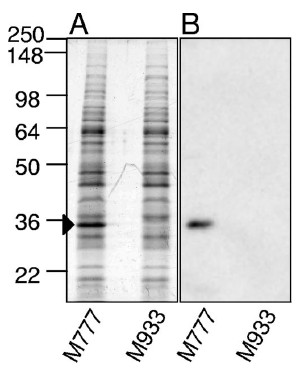
Although a lipL32 knockout mutant would help scientists figure out whether LipL32 plays an essential role in pathogenesis, targeted gene disruptions are extremely difficult with pathogenic Leptospira.  Fortunately, Ben Adler's group at Monash University obtained an insertion mutation in the lipL32 gene of L. interrogans by transposon mutagenesis.  This gave the Australians and their collaborators an opportunity to test the role of LipL32 in causing lethal infections in the hamster model of leptospirosis.  They first confirmed that the lipL32 mutant failed to express LipL32 by Western blotting the mutant with LipL32 antiserum.  I am showing the Coomassie-blue stained protein gel of the whole-cell lysate below so that you can appreciate the abundance of LipL32.  It is the most intensely stained band in the control L. interrogans strain, which has its lipL32 gene intact.

Whole-cell lysates of the L. interrogans lipL32 mutant (M933) and a control strain with the transposon in an intergenic region (M777) were run into SDS-acylamide gels and stained (panel A) or analyzed by Western blotting with LipL32 antiserum (panel B).  The M777 strain was demonstrated in an earlier study to be lethal for hamsters.  (Figure 1 from Murray et al., 2009.)

The survival curves show that the L. interrogans lipL32 mutant was just as lethal to hamsters as the parent L. interrogans with its lipL32 gene intact, irrespective of the infection route.  Hence, lipL32 is not necessary for lethal infections of hamsters, at least under the conditions used in this study.

Panel A: Groups of 8 hamsters were inoculated with 1,000 leptospires into the abdominal cavity.  Panel B:  Groups of 10 hamsters were inoculated with 106 leptospires dropped into the eye.  The slight difference in the survival curves was not statistically significant.  (Figure 5 from Murray et al., 2009)


Rats are the natural reservoir hosts of L. interrogans.  They can carry the spirochete for years in their kidney tubules without showing any signs of illness.  LipL32 could have a role in chronic infections.  The investigators therefore tested the ability of the lipL32 mutant to establish a chronic infection in laboratory rats.  They found that the lipL32 mutant (M933) was able to colonize the rat kidneys as well as the control M777 strain.  Kidneys from all 8 rats inoculated with the lipL32 mutant were culture positive.

At first glance it's surprising that lipL32 was not required for acute or chronic infection.  The authors pointed out that the function of LipL32 could be copied by other proteins found on the surface of Leptospira.  However, the study could have been strengthened by making two changes.  First, since the authors were trying to determine whether lipL32 was necessary for chronic infection, the rats should have been allowed to live for at least a few months before their kidneys were cultured.  Instead, the infection was allowed to proceed for only 15 days before the rats were sacrificed.  Second, they should have measured the bacterial load in the rat kidneys, either by plating serial dilutions of the kidney homogenates for colonies (although I don't know if this is feasible for Leptospira) or by quantitative PCR.  Clearly, more work needs to be done before anyone can conclude that LipL32 is not essential for chronic infection.


Featured paper

Murray G.L., Srikram, A., Hoke, D.E., Wunder Jr., E.A., Henry, R., Lo, M., Zhang, K., Sermswan, R.W., Ko, A.I., and Adler, B. (March 2009).  Major surface protein LipL32 is not required for either acute or chronic infection with Leptospira interrogans.  Infection and Immunity 77(3):952-958.  DOI: 10.1128/IAI.01370-08


Related posts

Saturday, July 24, 2010

Antigen presentation in the bloodstream: How invariant NKT cells are activated by Lyme disease spirochetes

The spirochete Borrelia burgdorferi is the tick-borne agent of Lyme disease, which affects the joints, nervous system, and heart.  After being deposited into the skin by an infected tick, the spirochete must enter the bloodstream so that it can circulate in the blood to gain access to its target organs.

The host doesn't sit idly as B. burgdorferi establishes an infection.  Invariant natural killer (iNKT) cells are one of the tools deployed by the immune system in its battle against the Lyme spirochetes.  Scientists know this because B. burgdorferi-infected mice lacking iNKT cells ended up with more spirochetes in their tissues and greater joint swelling than mice with a complete immune system.2

iNKT cells are an odd type of T cell.  Like other T cells, iNKT cells have a T cell receptor (TCR), yet they also express protein markers used to identify natural killer (NK) cells.  What makes the iNKT cell invariant is its TCR α chain, which comes only in the version dubbed Vα14 in mice and Vα24 in humans.  Even the β chain of the TCR of iNKT cells is restricted to three types in mice and just one in humans.  The lack of variation is unusual because the α and β TCR chains of conventional αβ T cells come in many forms in each individual, resulting in millions of varieties of TCRs.  This enables conventional αβ T cells to recognize a wide range of microbial peptide antigens when displayed by an MHC molecule on the surface of an antigen-presenting cell (see figure below).  In contrast, the TCRs of iNKT cells recognize a limited set of glycolipids displayed by the antigen-presenting cell's CD1d molecule, which structurally resembles MHC.  So far these glycolipids have been found only in Sphingomonas and B. burgdorferi.

Antigen recognition by T cells.  The "X" represents variable T cell receptor chains.
Figure 1 from ref. 3.
The structures of the B. burgdorferi glycolipids recognized by iNKT cells are shown below.  BbGL-IIc is recognized by mouse iNKT cells, and BbGL-IIf reacts with human iNKT cells.4
Structures of B. burgdorferi glycolipid antigens recognized by iNKT cells.  Figure 3d from ref. 3.

iNKT cells are activated when their TCR binds to BbGL-II complexed with CD1d.4  The activated iNKT cells secrete cytokines that elicit the appropriate immune response against the spirochetes.  How these cytokines promote killing of B. burgdorferi remains unknown.

To view the process of iNKT cell activation, scientists have recently obtained video footage of the early stages of the immune response to Borrelia burgdorferi circulating in the bloodstream of mice.1  The study by Lee et al., which appeared in the April issue of Nature Immunology, complements two earlier studies that revealed how the Lyme disease spirochete escapes from the bloodstream of mice to invade the surrounding tissues.5,6

The investigators employed fluorescence video microscopy to watch the immune cells in action following injection of an engineered B. burgdorferi strain expressing green fluorescent protein (GFP) into the bloodstream.  Although the spleen is better known for filtering bloodstream pathogens, the liver was selected for observation because iNKT cells make up 30% of the T cells in the liver.  In contrast, iNKT cells represent only 2.5% of T cells in the spleen.  Moreover, mice missing their spleen were able to limit B. burgdorferi infection as well as mice having a spleen, suggesting that the spleen is not critical in fighting bloodstream B. burgdorferi.

iNKT cells reside in the liver's sinusoids, which are the specialized capillaries that carry blood through the liver.  Similar to what other investigators have observed, the authors saw iNKT cells creeping along the inner surface of the liver sinusoids in healthy mice (see video below).

iNKT cells crawling within the liver sinusoids of a mouse genetically altered to express green fluorescent protein (GFP) in iNKT cells.  The iNKT cells glow bright green.  The elapsed time is shown at the top right.  Video 2 from ref. 1.

The investigators wanted to figure out which of the antigen-presenting cells found in the liver presented borrelial glycolipid to iNKT cells.  The answer?  After the spirochetes were injected into the bloodstream, they were quickly captured by Kupffer cells, the specialized blood-filtering macrophages that also reside in the liver sinusoids (see figure below).  Unlike iNKT cells, Kupffer cell remained stationary.

Capture of fluorescent B. burgdorferi (thin green bodies) by Kupffer cells (arrowhead).  Kupffer cells are stained red.  B. burgdorferi that avoided capture can be seen bound to the endothelium, trying to escape from the bloodstream into the liver tissue (arrow).  Figure 2e from ref. 1.

During the next several hours, the captured spirochetes were engulfed and broken up by the Kupffer cells so that BbGL-II could be loaded onto CD1d and displayed on the cell surface.  At 8 hours post injection, iNKT cells started to cluster and form stable contacts with Kupffer cells.  The iNKT cells were attracted to Kupffer cells churning out the chemokine CXCL9, a potent iNKT cell attractant.  The evidence for this was that injection of antibodies against the CXCL9 receptor, located on the iNKT cell surface, blocked clustering of iNKT cells.  Interaction of the Kupffer and iNKT cells was accompanied by increased blood and liver levels of the cytokine IFN-γ (interferon-gamma), a sign that the iNKT cells were being activated.

Left panel:  Liver 24 hours after injection of a GFP+ strain of B. burgdorferi into the bloodstream of a mouse.  Arrows indicate spirochetes (thin green bodies) that were not captured.  Kupffer cells are stained a red. The iNKT cells are the large bright green bodies.  The bright iNKT clusters overwhelm the faint red Kupffer cells, which are difficult to see.  Right panel:  To obtain a more convincing image showing contact between Kupffer cells and iNKT cells, a 3D reconstruction of the optical sections through the liver was performed.  Rotation of the image reveals interactions between Kupffer and iNKT cells.

Not all spirochetes were captured.  The investigators saw B. burgdorferi escaping from the sinusoids into the surrounding liver tissue even as other spirochetes were trapped by nearby Kupffer cells (see figures above).  Spirochetes circulating throughout the host probably escaped into other organs in the same manner.  Indeed, large amounts of  B. burgdorferi DNA were detected by PCR in several organs, including the liver, three days after the spirochetes were injected.  Although one doesn't usually think about the effects of Lyme disease on the liver, the authors pointed out that a mild hepatitis is common in Lyme disease patients.  In one prospective study, 40% of Lyme disease patients had at least one liver test abnormality.7

ResearchBlogging.orgBy now you may be wondering why the liver would devote such a high percentage of its T cells towards recognizing glycolipids that aren't found on most bacteria.  One answer is that microbes lacking the proper glycolipids may activate iNKT cells indirectly.3  For example, Salmonella typhimurium uses its LPS to coax antigen-presenting cells into making an endogenous glycolipid that gets presented to the iNKT cell by CD1d.8  It is also possible that glycolipids that are recognized by iNKT cells are present in other bacteria but are yet to be discovered.


Featured paper

1. Lee, W.Y., Moriarty, T.J., Wong, C.H.Y., Zhou, H., Strieter, R.M., van Rooijen, N., Chaconas, G., & Kubes, P. (2010). An intravascular immune response to Borrelia burgdorferi involves Kupffer cells and iNKT cells Nature Immunology, 11 (4), 295-302 DOI: 10.1038/ni.1855

Other references

2.  Tupin, E., Benhnia, M.R., Kinjo, Y., Patsey, R., Lena, C.J., Haller, M.C., Caimano, M.J., Imamura, M., Wong, C., Crotty, S., Radolf, J.D., Sellati, T.J., and Kronenberg, M. (2008).  NKT cells prevent chronic joint inflammation after infection with Borrelia burgdorferi.  Proc. Natl. Acad. Sci. USA 105(50):19863-19868.  DOI: 10.1073/pnas.0810519105

3.  Tupin, E., Kinjo, Y., and Kronenberg, M. (2007).  The unique role of natural killer T cells in the response to microorganisms.  Nature Reviews Microbiology 5(6):405-417.  DOI: 10.1038/nrmicro1657

4.  Kinjo, J., Tupin, E., Wu, D., Fujio, M., Garcia-Navarro, R., Benhnia, M. R., Zajonc, D.M., Ben-Menachem, G., Ainge, G.D., Painter, G.F., Khurana, A., Hoebe, K., Behar, S.M., Beutler, B., Wilson, I.A., Tsuji, M., Sellati, T.J., Wong, C., and Kronenberg, M. (2006).  Nature Immunology 7(9):978-986.  DOI: 10.1038/ni1380

5.  Moriarty, T.J., Norman, M.U., Colarusso, P., Bankhead, T., Kubes, P., and Chaconas, G. (June 20, 2008).  Real-time high resolution 3D imaging of the Lyme disease spirochete adhering to and escaping from the vasculature of a living host.  PLoS Pathogens 4(6):e1000090.  DOI: 10.1371/journal.ppat.1000090

6. Norman, M.U., Moriarty, T.J., Dresser, A.R., Millen, B., Kubes, P., and Chaconas, G. (October 3, 2008). Molecular mechanisms involved in vascular interactions of the Lyme disease pathogen in a living host.  PLoS Pathogens 4(10):e1000169.  DOI: 10.1371/journal.ppat.1000169

7.  Horowitz, H.W.,  Dworkin, B., Forseter, G., Nadelman, R.B., Connolly, C., Luciano, B.B., Nowakowski, J., O'Brien, T.A., Calmann, M., Wormser, G.P. (June 1996).  Liver function in early Lyme disease.  Hepatology 23(6):1412-1417.  DOI: 10.1002/hep.510230617

8.  Mattner J., DeBord, K.L., Ismail, N., Goff, R.D., Cantu III, C., Zhou, D., Saint Mezard, P., Wang, V., Gao, Y., Yin, N, Hoebe, K., Schneewind, O., Walker, D., Beutler, B., Teyton, L, Savage, P.B., and Bendelac, A. (March 24, 2005).  Exogenous and endogenous glycolipid antigens activate NKT cells during microbial infections.  Nature 434(7032):525-529.  DOI: 10.1038/nature03408

Related posts

Wednesday, March 31, 2010

A fresh approach towards a Lyme disease vaccine: targeting the tick

We tend to focus on the pathogen when thinking about how the immune system responds to tick-borne infections.  Ticks also provoke an immune response, even those that don't harbor any infectious agent.  Animals that are repeatedly bitten by ticks will eventually develop immunity to the tick.  There's even a commercial tick vaccine called TickGARD, which protects cattle against infestation by the tick Boophilus microplus.  TickGARD is formulated with a protein located in the midgut of B. microplus.

Animals that are immune to ticks also resist infection by some tick-borne pathogens, including Borrelia burgdorferi (Nazario et al., 1998).  Vaccines designed from a single tick component may also protect host animals from infectious agents.  A tick vaccine formulated with the tick cement protein 64TRP protected mice from being killed by the tick-borne encephalitis virus introduced by its vector, Ixodes ricinus (Labuda et al., 2006).

These earlier studies prompted Erol Fikrig's group at Yale to devise a vaccine that targets an Ixodes tick protein required by B. burgdorferi for a successful infection.  They focused on Salp15, a protein found in tick saliva.  Salp15 binds to the B. burgdorferi surface protein OspC as the spirochete passes through the salivary gland on its way into the skin of the victim.  Salp15 is one of the many bioactive salivary proteins that dampen the immune system to allow the tick to remain attached for several days so that it could complete its blood meal.  B. burgdorferi exploits Salp15 to fend off the immune response in the early stages of infection.  Since Salp15 coats B. burgdorferi, a vaccine targeting Salp15 could make B. burgdorferi vulnerable to killing by the immune system.  Indeed, Salp15 antiserum was able to enhance phagocytosis of Salp15-coated B. burgdorferi by mouse macrophages in vitro.

Despite the promising in vitro results, Salp15 as a vaccine was only partially protective in animal studies.  55-60% of mice actively immunized with Salp15 or passively immunized with Salp15 antiserum ended up infected with B. burgdorferi following challenge with infected Ixodes scapularis ticks.  Moreover, ticks were able to feed normally on mice immunized with Salp15, indicating that the animals did not acquire tick immunity.

Where Salp15 shined was in improving the efficacy of another Lyme vaccine.  The OspA vaccine requires several doses to achieve maximum protection against B. burgdorferi.  When Salp15 was combined with OspA, a single dose of the mixture spared 70% of mice from infection, whereas only 10-20% of mice immunized with a single dose of OspA or OspA plus Salp25D (an irrelevant tick salivary protein) were protected.  Thus future Lyme disease vaccines that target a component of the spirochete could also include Salp15 to enhance their protective capacity.

Scientists are undoubtedly examining other tick proteins as potential vaccines against ticks and the pathogens they transmit.

Featured paper

Dai, J., Wang, P., Adusumilli, S., Booth, C.J., Narasimhan, S., Anguita, J., and Fikrig, E. (November 19, 2009).  Antibodies against a tick protein, Salp15, protect mice from the Lyme disease agent.  Cell Host & Microbe 6:482-492.  DOI: 10.1016/j.chom.2009.10.006

Other references

Labuda, M., Trimnell, A.R., Licková, M., Kazimírová, M.,Davies, G.M., Lissina, O., Hails, R.S., and Nuttall, P.A. (April 2006).  An antivector vaccine protects against a lethal vector-borne pathogen.  PLoS Pathogens 2(4):e27.  DOI: 10.1371/journal.ppat.0020027

Nazario, S., Das, S., De Silva, A.M., Deponte, K., Marcantonio, N., Anderson, J.F., Fish, D., Fikrig, E., and Kantor, F.S. (June 1998).  Prevention of Borrelia burgdorferi transmission in guinea pigs by tick immunity.  American Journal of Tropical Medicine and Hygiene 58(6):780-785.  Link

Saturday, March 20, 2010

Tigecycline fails to eradicate persisting Borrelia burgdorferi

Antibiotics are usually successful in treating Lyme disease, especially if administered early.  The problem is that some patients continue to experience symptoms even after completing the recommended treatment regimen.  Although the current IDSA guidelines assert that the lingering symptoms are not due to persisting Borrelia burgdorferi,  the mouse model of Lyme disease clearly demonstrates the survival of live (albeit disabled) spirochetes following treatment with ceftriaxone, one of the antibiotics used to treat disseminated Lyme disease. As I wrote in an earlier post, the key question that must be answered is whether the lingering spirochetes are responsible for the persisting symptoms.  If so, a more potent antibiotic that could eliminate all of the spirochetes (or enough of them to allow the immune system to quickly mop up the rest) would be desired.

The newer antibiotic tigecycline was recently approved for treating skin and intra-abdominal infections caused by complex mixtures of bacteria.  Tigecycline is a tetracycline antibiotic, which blocks translation of mRNA into proteins by sticking tightly to the 30S ribosome subunit of bacteria.  In turns out that tigecycline exhibits greater antimicrobial activity than ceftriaxone (and doxycycline, another Lyme antibiotic) against B. burgdorferi, at least in the test tube.

Barthold and colleagues tested tigecycline to see if it could eradicate B. burgdorferi from persistently infected mice.  Groups of mice infected for 4 months with B. burgdorferi were treated with ceftriaxone (10 mice), a low dose of tigecycline (7 mice), or a high dose of tigecycline (9 mice).  A control group was sham treated with saline.  Three months after treatment was completed, the mice were examined to see if the spirochetes were still in the tissues.  As you might expect, B. burgdorferi DNA was detected by PCR at high levels in multiple tissues in all 10 mice that were administered saline, and the spirochetes were successfully cultured from the tissues.  In the ceftriaxone group, as shown in an earlier study by Barthold's lab, low levels of B. burgdorferi DNA were detected in leg joints from all 10 mice.  Although B. burgdorferi could not be cultured from the ceftriaxone-treated mice, ticks that fed on the mice were able to transmit the spirochetes to immunodeficient (SCID) mice, where B. burgdorferi was detected by PCR at the end of the experiment.  Since transmission requires active penetration of B. burgdorferi through several tissue barriers, the spirochetes that remained following antibiotic treatment must have been alive, although they could not be cultured.  Moreover, several B. burgdorferi mRNA transcripts were detected in some of the ceftriaxone-treated mice, another hint that the spirochetes remained viable (mRNA, unlike DNA, is extremely labile and would quickly degrade in dead bacteria).

How well did tigecycline work?  Despite its heightened potency against B. burgdorferi in test tube experiments and its much longer half-life in mice, tigecycline didn't work any better than ceftriaxone in eliminating the spirochetes, even at the higher dose.

The studies performed by Barthold's group raises several questions:
  • Would prolonging antibiotic treatment eventually eliminate the spirochetes?  Curiously, tigecycline was administered to the mice for only 10 days.
  • Do the spirochetes that remain following antibiotic treatment cause disease?  So far, the answer appears to be, "No."  Unlike the saline-treated mice, the antibiotic-treated mice did not exhibit any signs of disease.  Necropsies failed to reveal a inflammatory response against the spirochetes remaining in the tissues.
  • Do the spirochetes that survive antibiotic treatment give rise to disease later?  The earlier study by Barthold's group showed that although viable, the spirochetes were slowly diminishing in number in the tissues of mice that were treated with antibiotics.  If the mice were followed for a longer period of time, would the disabled spirochetes eventually disappear or revive to elicit a relapse of disease?
  • Is the mouse model even relevant to human Lyme disease?  Borrelia burgdorferi has evolved to persist in the mouse, its natural host, and may act differently in humans.  Obviously the experiments presented here can't be performed on humans, but an animal model that is more relevant to human Lyme disease may be more appropriate for addressing the issues raised by Barthold's work.
Featured paper

Barthold, S.W., Hodzic, E., Imai, D.M., Feng, S., Yang, X., and Luft, B.J. (February 2010).  Ineffectiveness of tigecycline against persistent Borrelia burgdorferi.  Antimicrobial Agents and Chemotherapy 54(2):643-651.  DOI: 10.1128/AAC.00788-09

Related paper

Hodzic, E., Feng, S., Holden, K., Freet, K.J., and Barthold, S.W. (May 2008).  Persistence of Borrelia burgdorferi following antibiotic treatment in mice.  Antimicrobial Agents and Chemotherapy 52(5):1728-1736.  DOI: 10.1128/AAC.01050-07

Thursday, August 20, 2009

Zebrafish model of leptospirosis: Where's the relevance?

Scrutinized for the past several decades as a model of embryonic development, the zebrafish has recently been promoted as a vertebrate model for investigating the pathogenesis of infectious diseases. Zebrafish embryos are transparent, allowing microbiologists to readily view the course of infections in real time. Another advantage of the zebrafish model is that it's amenable, at least in theory, to large-scale genetic screens for mutated host or microbial genes that affect the infection process.


Davis and colleagues observed the early stages of zebrafish embryo infection by the spirochete Leptospira interrogans, as described in a recent issue of PLoS Neglected Tropical Diseases. They injected 10-100 spirochetes into the hindbrain of zebrafish embryos at 30 hours post-fertilization, when the innate immune response is fully functional. Macrophages rushed to the hindbrain and engulfed the invaders within the first 4 hours following inoculation of the spirochetes. L. interrogans was also rapidly phagocytosed when injected into the caudal vein.

Figure 1a from Lesley and Ramakrishnan, 2008. A zebrafish embryo 30 hours following fertilization. The hindbrain and caudal vein are indicated with the bracket and arrow, respectively.

Macrophages typically kill and destroy their prey following phagocytosis. However, spirochetes were still observed in the macrophages 24 hours following inoculation, suggesting that L. interrogans can survive inside macrophages.

Their most striking observation was the accumulation of infected macrophages near the dorsal aorta in a region known as the aorta-gonad-mesonephros (AGM), where hematopoietic stem cells are born. This was not a general phenomenon of bacterial infections as macrophages harboring Pseudomonas aeruginosa failed to accumlate at the AGM following its injection into zebrafish embryos.

Figures 2C and 2D from Davis et al., 2009. The embryo was infected with fluorescently stained L. interrogans. 24 hours following infection, most of the spirochetes were found near the dorsal aorta (brackets), with a few scattered around the ventral tail. Scale bar, 300 µm in panel C, 100 µm in panel D.

Here's what the authors concluded in the final sentence of the paper:
The strikingly specific delivery of leptospires to [the AGM] by phagocytes provides insights into pathogenesis by suggesting a novel mechanism for targeting of organs during leptospiral dissemination.
In other words, L. interrogans may be capable of steering macrophages towards specific organs. Once the macrophages arrive at their destination, the spirochetes may escape from the macrophage and colonize the organ.

The challenge for the authors in future studies will be to demonstrate the relevance of the zebrafish model to leptospirosis. Hamsters and guinea pigs are appropriate models for leptospirosis because the pathology and lethality of Leptospira infection in these rodents is similar to what's observed in human leptospirosis patients. The fate of Leptospira that macrophages capture in these rodents differs from what is seen in zebrafish embryos. Leptospira that are found inside macrophages in tissue sections from infected rodents often appear to be disintegrating. Nevertheless, it's possible that a few Leptospira survive phagocytosis and subsequently guide the macrophage towards the target organs.

Featured paper

Davis, J.M., Haake, D.A., & Ramakrishnan, L. (2009). Leptospira interrogans stably infects zebrafish embryos, altering phagocyte behavior and homing to specific tissues PLoS Neglected Tropical Diseases, 3 (6) DOI: 10.1371/journal.pntd.0000463

Other references

Lesley, R. and Ramakrishnan, L. (2008). Insights into early mycobacterial pathogenesis from the zebrafish. Current Opinions in Microbiology 11(3):277-283. DOI: 10.1016/j.mib.2008.05.013

Thursday, May 28, 2009

Leptospira heme oxygenase frees iron from heme

ResearchBlogging.orgI mentioned in a recent post that iron is an essential trace metal that bacteria must acquire from its surroundings. (From that same post you will also recall that the Lyme disease spirochete B. burgdorferi is a rare exception that doesn't need iron.) Much of the iron in our body is trapped within the center of the heme molecule. Heme itself is not readily accessible as it is bound to host proteins such as hemoglobin. Pathogenic bacteria have evolved sophisticated systems to kidnap heme from host proteins and transport them into the cytoplasm. These complex systems, which include secreted degradative enzymes, heme capturing proteins, and transporter proteins that sit in the membrane, have been examined in numerous bacteria. However, the fate of heme after it is acquired by the bacteria is poorly understood. In some cases, the captured heme may be incorporated into bacterial proteins such as cytochromes, which participate in electron transport. In other cases, bacteria may need to extract the iron trapped in the middle of the heme molecule. Some bacteria possess the enzyme heme oxygenase, which extracts the iron caged within heme by the following reaction (adapted from Scheme 1 in Kikuchi et al., 2005):
Unlike its cousin that causes Lyme disease, the spirochete Leptospira requires iron for growth. Ben Adler's group at the Monash University in Australia isolated a Leptospira interrogans mutant with the transposon TnSC189 inserted into hemO, the gene encoding heme oxygenase. The properties of the hemO mutant is described in two papers in the journal Microbes and Infection. The graph below (figure 1B in Murray et al., 2008) shows that growth of the hemO mutant is impaired, although not completely, when hemoglobin is the sole source of iron in the culture medium. The residual growth of the mutant indicates that L. interrogans may possess another activity that extracts iron from heme.
In their follow-up study, Adler's group demonstrated that the hemO gene was necessary for L. interrogans to fully express its virulence in the hamster model of leptospirosis. For this study they used the hemO mutant and a control L. interrogans strain that had the TnSC189 element inserted in a noncoding region, presumably where gene expression would not be affected. The two strains were injected into the abdominal cavities of separate groups of hamsters, which were then monitored for 14 days. Only 8 of 24 hamsters (33%) survived the challenge with the control strain, whereas 20 of 24 (83%) injected with the hemO mutant survived. The difference in survival rates between the two groups was statistically significant (P = 0.001).

Although the hemO mutant was ineffective at killing hamsters, it was still able to colonize the kidneys of most of the animals. Colonization was assessed by culturing kidney or urine in Leptospira growth medium. The mutant was recovered by culturing of kidney or urine from 17 of 20 hamsters that survived the challenge with the hemO mutant and all 3 that died. These results were similar to what was obtained with hamsters inoculated with the control strain, which was recovered from all 8 animals that survived and all 12 that died. (Not all hamsters were examined for colonization.)

Why was the hemO mutant able to colonize the kidney when it was unable to extract iron from heme? Heme is not the only source of iron in the body. The mutant may have captured one of the other forms of iron present in the host. The genome of L. interrogans encodes several homologs of transporters that the spirochete may use to acquire non-heme sources of iron (Louvel et al., 2006). Since these other iron sources are less abundant than heme, the tissue burden (density of bacteria) of the mutant in the kidneys may have been lower than that of the control strain thereby allowing most of the hamsters challenged with the hemO mutant to survive.

One obvious limitation of the study is that the investigators did not attempt to complement the hemO mutation with a wild-type copy of the gene. However, I should point out that currently no plasmid is available that replicates in L. interrogans, rendering complementation of L. interrogans mutations difficult. The researchers did verify that the gene immediately downstream of hemO was still transcribed in the mutant.

This work is significant for the following reasons. First, although there have been two other studies that have examined the role of Leptospira genes in virulence, this study was the most satisfying to read because it was the first to show that a gene encoding a product of known function has a role in the virulence of Leptospira. Second and perhaps more importantly, it is the first to demonstrate the importance of a bacterial heme oxygenase in virulence.

Featured papers

Murray, G., Ellis, K., Lo, M., & Adler, B. (2008). Leptospira interrogans requires a functional heme oxygenase to scavenge iron from hemoglobin Microbes and Infection, 10 (7), 791-797 DOI: 10.1016/j.micinf.2008.04.010

Murray, G., Srikram, A., Henry, R., Puapairoj, A., Sermswan, R., & Adler, B. (2009). Leptospira interrogans requires heme oxygenase for disease pathogenesis Microbes and Infection, 11 (2), 311-314 DOI: 10.1016/j.micinf.2008.11.014

Other references

Kikuchi, G., Yoshida, T., and Noguchi, M. (2005). Heme oxygenase and heme degradation. Biochemical and Biophysical Research Communications 338(1):558-567. DOI: 10.1016/j.bbrc.2005.08.020

Louvel, H., Bommezzadri S., Zidane, N., Boursaux-Eude, C., Creno, S., Magnier, A., Rouy, Z., Médigue, C., Saint Girons, I., Bouchier, C., and Picardeau, M. (2006). Comparative and functional genomic analyses of iron transport and regulation in Leptospira spp. Journal of Bacteriology 188(22):7893-7904. DOI: 10.1128/JB00711-06

Thursday, April 30, 2009

The manganese transporter of the iron-free Lyme disease spirochete is essential for infection

ResearchBlogging.orgIron is a trace element essential for life. As such, living beings, including humans, must obtain iron from their diet. The same holds true for the bacteria that make us sick. Unfortunately for bacteria, iron is not readily available as it is tied up by various proteins in our body. Consequently, pathogenic bacteria have devised clever strategies to wrest iron away from these proteins.

The Lyme disease spirochete Borrelia burgdorferi lacks the machinery necessary for acquiring iron. In fact, B. burgdorferi can grow in the absence of iron. The reason that B. burgdorferi does not need iron is that the spirochete is missing most of the common metalloproteins that require iron to function. For the few metalloproteins that are present, manganese (Mn) may substitute for iron. This raises the question of how B. burgdorferi acquires Mn.

In the March 3 issue of PNAS, Ouyang and colleagues describe a potential Mn transporter BmtA (Borrelia metal transport protein A) encoded in the B. burgdorferi genome. What is their evidence that BmtA is a Mn transporter?
  • Analysis of its amino acid sequence indicates that BmtA is a member of the ZIP family of metal transporters. The transporters sit in the membrane and transport metals such as iron, zinc, and manganese across the membrane. Most ZIP family members are predicted to have 8 transmembrane domains with potential metal-binding histidine residues within a "variable region" (see figure below taken from Guerinot 2000) and a signature sequence in the fourth transmembrane domain containing another metal binding histidine. BmtA lacks the his-X-his-X-his (X = any amino acid) metal binding site in the variable region, but it does have the signature sequence.
  • When the bmtA gene was knocked out, the mutant B. burgdorferi was still able to grow in culture, but it was unable to accumulate Mn in its cytoplasm. The ability to accumulate Mn was restored when a plasmid expressing bmtA was introduced into the mutant.

To show conclusively that BmtA is a Mn transporter, the authors will need to incorporate purified BmtA into an artificial lipid bilayer (liposome) and demonstrate transport of Mn across the membrane.

The investigators next determined whether BmtA was required for infection of the mouse model of Lyme disease. They found that the bmtA mutant was unable to infect mice when injected into the skin. Infection was assessed by culturing heart, joint, and skin tissue removed 4 weeks after inoculation. The effect of the knockout was striking. None of the 66 organs sampled from the 22 mice inoculated with the bmtA mutant were culture positive. On the other hand, all 21 organs obtained from the 7 mice injected with the wild-type B. burgdorferi strain were culture positive. Infectivity was restored when the bmtA gene was introduced on a plasmid back into the bmtA mutant.

Because the bmtA knockout mutant grew in vitro yet was unable to grow in mice, BmtA must be essential for infectivity. Ouyang et al. presented two possible roles of BmtA in virulence in the Discussion of their paper. First, BmtA may be required for the activity of superoxide dismutase, which in B. burgdorferi is predicted to require Mn rather than iron. Superoxide dismutase detoxifies the reactive oxygen species (ROS) generated by phagocytic cells (neutrophils and macrophages) trying to ward off invading bacteria. Indeed, Ouyang et al. demonstrated that knocking out the bmtA gene rendered B. burgdorferi more sensitive to the oxidizing agent t-butyl hydroperoxide.

Second, Mn may contribute to the regulation of Borrelia genes encoding virulence determinants. The authors present as an example the transcriptional regulator BosR, which they state is a "Mn-dependent Fur homolog." This is incorrect as two different research groups have shown Mn to have no effect or even inhibit the activity of BosR. Nevertheless, there may be other Mn-dependent regulators of borrelial gene expression yet to be discovered.

The authors tout BmtA as a discovery that "may lead to new strategies for thwarting Lyme disease." That's probably true, but a word of caution should be expressed here. Any inhibitor of BmtA that's identified in future studies must have high specificity for the borrelial protein since ZIP family proteins are also found in humans.

Featured paper

Ouyang, Z., He, M., Oman, T., Yang, X., & Norgard, M. (2009). A manganese transporter, BB0219 (BmtA), is required for virulence by the Lyme disease spirochete, Borrelia burgdorferi Proceedings of the National Academy of Sciences, 106 (9), 3449-3454 DOI: 10.1073/pnas.0812999106

Other references

Boylan, J.A., Posey, J.E., and Gherardini, F.C. (2003). Borrelia oxidative stress response regulator, BosR: A distinctive Zn-dependent transcriptional activator. Proceedings of the National Academy of Sciences USA 100(20):11684-11689.

Guerinot M.L. (2000). The ZIP family of metal transporters. Biochimica et Biophysica Acta 1465(1-2):190-198.

Posey, J.E. and Gherardini, F.C. (2000). Lack of a role for iron in the Lyme disease pathogen. Science 288(5471):1651-1653.

Tuesday, March 10, 2009

Rhesus monkeys with Lyme spirochetes in the brain

ResearchBlogging.orgLyme disease has been mentioned in the news a lot lately. First it was a pet chimp mauling his neighbor last month in a Connecticut town. Many news accounts of the assault reported that Travis the chimp suffered from Lyme disease and had been given tea laced with Xanax to calm his erratic behavior prior to the attack. Investigators may never figure out what provoked Travis to attack Charla Nash. Was Lyme disease a factor? Maybe (although if I had to guess, it had more to do with a wild animal being kept as a pet, living where it didn't belong).

This past weekend a human reportedly suffering from long-term Lyme disease gunned down a pastor and injured several parishioners in an Illinois church. As with any topic having to do with chronic Lyme disease, experts disagree on whether Lyme disease can trigger aggression (but I'm pretty sure what the shooter's defense attorney believes). One prominent researcher even questions whether the alleged shooter truly had Lyme disease.

A blogger on Forbes.com doubts whether chimps could be stricken with Lyme disease. In reality, seeing a chimp with Lyme disease should not surprise us at all since another nonhuman primate, the rhesus monkey, has been used for years to study neuroborreliosis, the form of Lyme disease caused by infection of the nervous system. Neuroborreliosis is observed in up to 15% of untreated Lyme disease patients. I will present two studies in which scientists examined infection of the central nervous system of rhesus monkeys by the Lyme disease spirochete, Borrelia burgdorferi.

In a 2000 study by Cadavid and colleagues, the investigators found small numbers of spirochetes in the meninges and spinal cord nerve roots of rhesus monkeys four months following laboratory infection with B. burgdorferi by needle inoculation into the skin (see figure below). In humans, the inflammatory response to the spirochetes in the meninges and nerve roots can cause meningitis (headache, stiff neck, and sensitivity to light) and radiculoneuritis (shooting pains and abnormal skin sensations), respectively.

Figure 2b from Cadavid et al., 2000. A single B. burgdorferi cell in the anterior nerve root stained by immunohistochemistry with anti-B. burgdorferi antibodies.

The investigators found no convincing evidence for the presence of B. burgdorferi in the brain parenchyma of the infected rhesus monkeys. B. burgdorferi was not found in the brain by microscopy. On the other hand, B. burgdorferi DNA was detected in brain tissue by polymerase chain reaction (PCR). This result may suggest that the spirochete is present in the brain at low levels, although the authors believed that spirochetes in the meninges surrounding the brain was the real source of the DNA. It's also possible that the N40 B. burgdorferi strain used by the authors was incapable of invading the brain of rhesus monkeys and that another strain if injected into the skin would have found its way to the brain.

Two conditions associated with neuroborreliosis disrupt brain function in humans. The first is Lyme encephalopathy, which can cause fatigue and problems with concentration and memory. Brain activity is clearly affected in patients with Lyme encephalopathy, yet the underlying pathogenic mechanism remains an enigma. Many patients with this potentially disabling condition do not exhibit the classic signs of central nervous system inflammation such as the presence of white blood cells in their cerebral spinal fluid (CSF), production of antibodies against the infective agent in the CSF, or abnormal brain MRIs. Some neuroborreliosis experts have proposed that these cases of Lyme encephalopathy are a result of "toxic-metabolic" effects of infection elsewhere in the body (such as Lyme arthritis in the joints) spilling into the brain. Nevertheless, a low-level infection of the brain by B. burgdorferi cannot be ruled out. More obvious brain involvement is observed in neuroborreliosis patients with encephalitis, in whom brain lesions are detected by MRI, accompanied by white blood cells and production of anti-B. burgdorferi antibodies in the CSF. Symptoms include slight weakness or paralysis affecting one side of the body, spastic muscles, inability to feel sensations, and bladder dysfunction. In rare cases, patients may experience strokes or seizures. Patients with encephalitis are likely to have spirochetes in their brain, usually near blood vessels.

Lyme disease may also be associated with psychiatric illness, particularly depression. Rare cases of panic attacks, bipolar disorder, mania, obsessive-compulsive disorder, dementia, and violent outbursts have also been reported.

A recent study by Mario Philipp's group in Tulane, published recently in The American Journal of Pathology, described the potential effects of B. burgdorferi on brain cell activity of rhesus monkeys. Spirochetes injected into the skin with a needle or by tick bite are unable to make it into the brain of rhesus monkeys, as I explained above. Therefore, the investigators injected live B. burgdorferi spirochetes directly into the right side of the brains of rhesus monkeys. After two weeks, they found that an average of ~10% of oligodendrocytes at the injection sites had undergone death by apoptosis, as detected by fluorescent TUNEL staining (see figure below). In contrast, fewer than 2% of oligodendrocytes underwent apoptosis when sites in the left side of the brain of the same animals were injected with saline. Oligodendrocytes produce the myelin sheaths that surround the axons of neurons and promote transmission of electric impulses; one would expect the loss of oligodendrocytes to affect neuron function.

The in vivo results corroborated ex vivo experiments in which brain slices from rhesus monkeys were incubated with B. burgdorferi for up to eight hours. In the ex vivo experiments, apoptosis of both oligodendrocytes and neurons were detected by fluorescent TUNEL staining and with antibody against activated caspase 3, a more specific marker of apoptosis. The authors speculated that neurons undergoing apoptosis in vivo were removed by phagocytes and therefore went undetected in the brain injection experiment. The other major brain cell types, microglial cells and astrocytes, were spared from apoptotic death both ex vivo and in vivo.

Figure 4A from Ramesh et al., 2008. Oligodendrocytes are stained red with antibody to S-100. Nuclei of cells undergoing DNA fragmentation, a consequence of apoptosis, are TUNEL stained green. When the images are merged, the nuclei of oligodendrocytes undergoing apoptosis become yellow. B. burgdorferi, which are stained in blue, do not appear to be in physical contact with the oligodendrocytes.

Figure 4B from Ramesh et al., 2008. Percentage of oligodendrocytes undergoing apoptosis from 3 sites of spirochete (A-C) and saline (D-F) injections (click on image for larger view).

The Tulane group also detected myriad cytokines and chemokines such as IL-6, IL-1ß, and CXCL13 being produced in the B. burgdorferi-treated brain slices by immunofluorescence antibody staining and DNA microarray analysis. The authors concluded that B. burgdorferi invasion of the brain elicits production of a brew of inflammatory mediators that promote apoptotic death of oligodendrocytes and neurons. It may be true that brain invasion by B. burgdorferi in cases of human neuroborreliosis leads to production of these cytokines and chemokines, as IL-6, IL-1ß, and CXCL13 have been detected in the CSF of neuroborreliosis patients in earlier studies. However, the apoptotic cells observed by the Tulane group may be the unnatural consequence of small areas of the brain being exposed to extremely large numbers of spirochetes. Each site was injected with 5,000 spirochetes, a number far greater than the rare spirochetes observed by microscopy in the brains of neuroborreliosis patients. For the ex vivo experiments, a 2-mm section of brain was incubated with 20 million spirochetes, again a large burden of bacteria. More convincing would be finding apoptosis of oligodendrocytes and neurons in brain tissues in patients with Lyme encephalitis. Clearly more studies are needed to understand the events that really occur when B. burgdorferi invades the brain.

References

Ramesh, G., Borda, J., Dufour, J., Kaushal, D., Ramamoorthy, R., Lackner, A., & Philipp, M. (2008). Interaction of the Lyme Disease Spirochete Borrelia burgdorferi with Brain Parenchyma Elicits Inflammatory Mediators from Glial Cells as Well as Glial and Neuronal Apoptosis American Journal Of Pathology, 173 (5), 1415-1427 DOI: 10.2353/ajpath.2008.080483

Diego Cadavid, Tim O'Neill, Henry Schaefer, and Andrew R. Pachner (2000). Localization of Borrelia burgdorferi in the Nervous System and Other Organs in a Nonhuman Primate Model of Lyme Disease Laboratory Investigation, 80 (7), 1043-1054

Saturday, February 7, 2009

The Lyme disease spirochete hijacks fibronectin to escape from the bloodstream

ResearchBlogging.orgThe glycoprotein fibronectin is a component of the molecular mesh known as the extracellular matrix, which not only provides physical support for our cells but also directs cellular activities during embryonic development, tissue repair, and other processes. High levels of soluble fibronectin (300 μg/ml) are also found in our bloodstream, where it quietly circulates until it is recruited to stabilize clots and promote wound repair.

Fibronectin has a modular organization consisting of binding sites for various matrix and cell surface molecules. Examples include attachment sites for integrins (labeled "Cell" in the figure below) and glycosaminoglycans (labeled "Heparin"), which are exposed on the surface of the endothelial cells that line our blood vessels.


B. burgdorferi is injected into the skin by an infected tick and spreads outward within the dermis, causing the familiar "bulls-eye" rash in some Lyme disease patients. The spirochete may eventually enter the bloodstream so that it can spread to other tissues. While in the bloodstream, the spirochete is surrounded by fibronectin. In fact, B. burgdorferi attaches to fibronectin in vitro, suggesting a role for fibronectin in Lyme disease.

It turns out that B. burgdorferi exploits the adhesive properties of plasma fibronectin to bind to the vessel wall before escaping into the surrounding tissue. Like their earlier work, which I described in my last post, this follow-up study by Norman and colleagues was conducted with fluorescent B. burgdorferi injected into the veins of live mice. The interactions of the spirochete with the capillary wall were observed by fluorescent intravital microscopy. The earlier study revealed that most of the interactions were transient, lasting for less than a second. B. burgdorferi was also seen crawling (dragging) along the vessel wall, which was followed by escape into the tissue or by stationary adhesion, a more intimate association with the vessel wall. Stationary adhesion could also be followed by extravasation and escape of the spirochete into the tissue. Both stationary adhesion and escape usually occurred between the endothelial cells lining the vessel wall. Each type of interaction (transient, dragging, and stationary adhesion) was quantitated by counting.

In their follow-up study, the authors demonstrated that coinjection of anti-fibronectin antibody and B. burgdorferi into the bloodstream of the mice diminished all catagories of interactions (transient, dragging, and stationary adhesion) by at least 90%. Control antibody (goat IgG) had no effect. These results indicate that fibronectin has a key role in mediating the attachment of B. burgdorferi to the microvasculature. Since fibronectin could potentially bind to GAGs and integrins on endothelial cells, the investigators also coinjected B. burgdorferi with peptides or antibodies known to block attachment of fibronectin to these targets. They found that the GAG-specific peptide reduced the interaction of B. burgdorferi with the vessel wall, whereas the integrin-specific peptide and antibodies had little effect. Thus, fibronectin may serve as a molecular bridge linking B. burgdorferi to GAGs displayed on the endothelial cells lining the blood vessel.

Which B. burgdorferi factor is involved in adherence to the vessel wall in vivo? Past studies had shown that the borrelial protein BBK32, a known fibronectin binding protein, mediated attachment of B. burgdorferi to fibronectin in vitro. Therefore, BBK32 was a logical candidate. To determine whether BBK32 was involved in vascular interactions in the mouse model, the research team employed a noninfectious B. burgdorferi strain that had lost bbk32 and other genes during long-term culture. The noninfectious strain failed to interact with the vasculature in the mouse. However, expression of BBK32 restored the ability of the noninfectious strain to transiently interact and drag along the vessel wall but only partly restored stationary adhesion. This results suggest that although BBK32 plays a role in vascular adherence, other bacterial factors are also involved.

Many pathogens have been shown to interact with fibronectin and GAGs in vitro. However, this study is highly significant as it is the first to demonstrate a role for these host molecules in bacterial adherence to the microvasculature in a living animal. Other spirochetes such as Treponema pallidum and Leptospira also express fibronectin binding proteins. Hence, the mechanism employed by B. burgdorferi to escape from the bloodstream may be similar for all disease-causing spirochetes. Moreover, other microbial pathogens have been shown to stick to fibronectin and GAGs in vitro. Thus, a large number of invasive pathogens may employ similar mechanisms to spread to different tissues via the bloodstream.

M. Ursula Norman, Tara J. Moriarty, Ashley R. Dresser, Brandie Millen, Paul Kubes, George Chaconas (2008). Molecular Mechanisms Involved in Vascular Interactions of the Lyme Disease Pathogen in a Living Host PLoS Pathogens, 4 (10) DOI: 10.1371/journal.ppat.1000169