Tuesday, February 15, 2011

The quest for outer membrane proteins of the stealth pathogen Treponema pallidum: the cliffhanger episode

Syphilis patients are able to generate antibodies against the spirochete Treponema pallidum.  However, if you were to mix sera from these patients with T. pallidum in a test tube, very few of the antibodies in the sera would bind to the spirochetes.  The reason is that the strange outer membrane architecture of T. pallidum makes the spirochete invisible to the antibodies.  Most of the proteins and lipid molecules targeted by the antibodies lie beneath the outer membrane.

The outer membrane of T. pallidum differs considerably from that of a typical Gram-negative bacterium.  The most glaring difference is that T. pallidum lacks lipopolysaccharide (LPS), a favorite target of the immune response.  The outer membrane is also bare of other potential surface antigens except for a very small number of transmembrane outer membrane proteins (Omps) and (possibly) surface lipoproteins.  The poor surface antigencity may help the so-called "stealth pathogen" persist in the body despite a robust immune response to the infection.

The few Omps displayed on the surface of T. pallidum must be doing something really important if the spirochete is willing to risk exposing them to attack by antibodies.  For this reason, scientists have been seeking the identity of these Omps to figure out what they do.  These rare Omps could also be fashioned into a long-desired syphilis vaccine.

Unfortunately, the scarcity of Omps and the delicate nature of the outer membrane of T. pallidum have stymied efforts to identify Omps.  The routine centrifugation and washing steps used to prepare other bacteria for analysis easily damage the outer membrane of T. pallidum, causing the loss of Omps and exposing the abundant periplasmic and inner membrane proteins.  Consequently, probes used to identify exposed proteins may react with the periplasmic and inner membrane proteins, which are normally shielded by the outer membrane.  Without the proper controls, this would lead one to conclude wrongly that a non-Omp that reacts with the probe (such as antibodies raised against the protein of interest) is surface exposed.  In addition, when the outer membrane is purified with the intention to identifying Omps, it is hard to distinguish the tiny amounts of Omps from proteins from other bacterial compartments contaminanting the outer membrane preparation.

Despite these technical challenges, several Omp candidates have been proposed, but those proteins are either mired in controversy (TprK, for example) or await experimental confirmation of their surface exposure.  To date, no protein that has been demonstrated unambiguously to be displayed on the exterior of T. pallidum.

The past decade has seen the development of several computer programs that can be used to predict whether a given gene encodes a transmembrane Omp.  The algorithms differ, but all of these programs attempt to identify amino acid sequences that fold into a β-barrel, which forms the core of transmembrane Omps whose 3D structures are known.  The β-barrel forms when an anti-parallel β-sheet rolls into the shape of a barrel.  Most of the β-barrel is embedded in the outer membrane so that the loops connecting the β-strands stick out from the two surfaces of the membrane.  The loops displayed on the external face of the outer membrane would be accessible to antibodies.  The size and composition of the loops vary among different Omps, but all β-strands tend to have alternating hydrophobic amino acids with their nonpolar side chains acid protruding out from the barrel into the hydrophobic interior of the lipid bilayer.  Each β-strand consists of 9-11 amino acid residues and is tilted up to 45° out of the transmembrane axis.  Different β-barrels have as few as 8 and as many as 22 transmembrane β-strands.

The ribbon representation of OmpA, an 8-stranded transmembrane Omps from E. coli, is shown below as one example.

From Figure 1b of Smith et al., 2007.  The N- and C-terminal β-strands are colored brown and blue, respectively.  The side chains of the "aromatic girdle" are shown.
Below, OmpA is unfurled to show the topology of the protein:

From Figure 1a of Smith et al., 2007.  β-strand amino acid residues are depicted as diamonds, and loop residues are depicted as circles.  Alternating hydrophobic amino acid residues within the  β-strand are colored red (aromatic residues of the girdle) and yellow.
Assuming that the rare transmembrane Omps of T. pallidum share the β-barrel structure, the obvious computational approach to finding these Omps would be to run all of the proteins encoded by the T. pallidum genome through one of these programs.  One problem with these programs is that they will pick up a few proteins that are not truly transmembrane Omps.  To minimize this problem, Justin Radolf's group, as reported in the December 2010 issue of Infection and Immunity, ran the 1038 protein-coding sequences of T. pallidum through seven different Omp-predicting programs.  They found that two proteins were predicted by all seven programs to have the β-barrel structure; another four candidates were identified by six programs.

One of the proteins at the top of the list, identified by all seven programs, was TP0326, a BamA homolog encoded by the genomes of many Gram-negative bacteria.  Experiments with other bacteria have shown that BamA is a member of an outer membrane protein complex that assembles other transmembrane Omps into the outer membrane, so it would make sense for T. pallidum to possess such a protein.  BamA itself is thought to be a transmembrane Omp. 

This wasn't the first time that a syphilis researcher has encountered TP0326.  In a study published 11 years ago, before the function of BamA was known, Caroline Cameron and colleagues demonstrated that antibodies raised against TP0326 (also called "Tp92" in their paper) stimulated macrophages to engulf T. pallidum in a process called opsonophagocytosis.  In addition, TP0326 was somewhat effective as a vaccine in the rabbit model of syphilis:  rabbits that had been immunized with TP0326 experienced milder skin lesions than unimmunized rabbits following inoculation of T. pallidum into the skin.  These observations indirectly supported the localization of TP0326 to the outer membrane since opsonophagocytosis and effective vaccination require a target that is accessible on the surface of the spirochete.  However, this earlier work lacked a more direct test such as the indirect immunofluorescence assay to confirm that TP0326 was exposed on the surface.

The problem with the standard two-step indirect immunofluorescence assay is that it is not sensitive enough to detect the rare Omps of T. pallidum.  Therefore, as described in the Infection and Immunity paper, Radolf's group tinkered with the assay and managed to amplify the output signal by adding a third step to the procedure.  To minimize damage to the outer membrane during the centrifugation and washing steps, the spirochetes were encased in gel microdroplets, which protected the delicate outer membrane while allowing antibodies to permeate to probe the T. pallidum surface.

With the modified immunofluorescence assay, the investigators were able to detect surface proteins with syphilitic antibodies for the first time, although only in a small minority of the spirochetes in the field of view lit up with the red color (see figure below).  Presumably, the other spirochetes failed to react with the antibodies because they didn't quite have enough Omp antigens being expressed on their surface (although a more interesting explanation would be that the nonreactive spirochetes had down-regulated their surface Omps).  Regardless of the true explanation, these results indicated that at least some of the antibodies generated by syphilis patients were directed against surface components of T. pallidum.  When the investigators treated the spirochetes with the detergent Triton X100 to intentionally damage the outer membrane, all of the spirochetes glowed, indicating that most of the antibodies targeted proteins beneath the surface of T. pallidum.  As a negative control, they demonstrated that sera from healthy patients failed to react with intact spirochetes.

To keep track of how many spirochetes were damaged by the procedure, the investigators added antibody raised against the periplasmic flagella along with the patient antibodies.  The flagellar antibodies would bind to the spirochetes only if the integrity of the outer membrane was compromised by handling the spirochetes.  The assay was designed so that bound flagellar antibodies would glow green.

From Figure 4 of Cox et al., 2010.   (A) All spirochetes, whether or not they fluoresced, could be seen with darkfield optics (DF).  Spirochetes that bound to antibodies from syphilis patients (HSS) glowed red.   Spirochetes with a disrupted outer membrane reacted with the flagellar antibody (anti-FlaA) and glowed green.  (B) 5.8% of the spirochetes observed were undamaged and reacted with patient antibodies (glowed red but not green).  Another 5.1% were damaged (glowed red and green).  89.0% of the spirochetes failed react with the patient antibodies.  100% of the spirochetes fluoresced when treated with the detergent Triton X100 before adding the antibodies.

With an improved immunofluorescence assay, the investigators were poised to test the proteins at the top of the list for surface exposure.  As I was nearing the end of the paper, I was expecting the authors to describe their test of the BamA homolog TP0326 for surface exposure.  Surprisingly, they ended the paper without testing any of the proteins near the top of the list.

I can only assume that the authors are planning to submit a separate manuscript in the future describing the successful detection of TP0326 or another protein near the top of the list.  But the problem with ending the paper without demonstrating surface localization of even a single protein is that one can question whether even the 3-step immunofluorescence assay is sensitive enough to detect an Omp exposed on the T. pallidum surface.  They did test two proteins lower down on the list that other labs believe are surface exposed (TprK and a fibronectin-binding lipoprotein)  but neither protein was detected on the outer membrane surface by the modified immunofluorescence assay.  So we are left with an assay that certainly has more sensitivity, but is it sensitive enough?

To be continued...(?)

Featured paper

Cox, D.L., Luthra, A., Dunham-Ems, S., Desrosiers, D.C., Salazar, J.C., Caimano, M.J.., and Radolf, J.D. (December 2010).  Surface immunolabeling and consensus computational framework to identify candidate rare outer membrane proteins of Treponema pallidumInfection and Immunity 78(12):5178-5194.  DOI: 10.1128/IAI.00834-10

Other references

Radolf, J.D. (June 1995).  Treponema pallidum and the quest for outer membrane proteins.  Molecular Microbiology 16(6):1067-1073.

Cameron, C.E., Lukehart, S.A., Castro, C., Molini, B., Godornes, C., and Van Voorhis, W.C. (April 2000).  Opsonic potential, protective capacity, and sequence conservation of the Treponema pallidum subspecies pallidum Tp92.  Journal of Infectious Diseases 181(4):1401-1413.  DOI: 10.1086/315399

Cox, D.L., Akins, D.R., Porcella, S.F., Norgard, M.V., and Radolf, J.D. (1995).  Treponema pallidum in gel microdroplets:  a novel strategy for investigation of treponemal molecular architecture.  Molecular Microbiology 15(6):1151-1164.

Image source

Smith, S.G.J, Mahon, V., Lambert, M.A., and Fagan, R.P. (August 2007).  A molecular Swiss army knife:  OmpA structure, function and expression.  FEMS Microbiology Letters 273(1):1-11.  DOI: 10.1111/j.1574-6968.2007.00778.x

Saturday, January 1, 2011

Designing a Lyme disease vaccine to attack the tick vector

Conventional vaccines target the surface components or secreted toxins of pathogens.  Erol Fikrig's group at Yale University has been exploring an unconventional approach towards developing a vaccine for Lyme disease, which is caused by a tick-borne pathogen.  Their recent work, published in the November issue of PLoS Pathogens, demonstrated partial success in protecting laboratory mice by immunization with a protein found in the saliva of the Ixodes tick vector.

Ixodes ticks spend several days feeding on blood while attached to the victim's skin.  B. burgdorferi is carried into the victim's skin in the Ixodes tick's saliva starting 3-4 days (on average) after attachment.  Tick saliva contains a blend of biological substances that aid the tick in drinking blood from its victim.  These substances include cement proteins to keep the tick's feeding apparatus tightly bound to the skin, anti-coagulants to keep the blood flowing into the tick, and anti-inflammatory factors that ward off the local inflammatory response.  The activity of these substances also promote transmission of B. burgdorferi from the feeding tick to the victim.  Hence a vaccine that targets a saliva component may protect humans from Lyme disease.

A Lyme vaccine that targets the tick has a few advantages over one that targets the spirochete.  First, a tick-based Lyme disease vaccine is unlikely to interfere with laboratory diagnosis, which currently relies on detection of antibodies against the Lyme Borrelia spirochete.  Second, an effective vaccine that targets the tick may also prevent transmission of other pathogens carried by the Ixodes tick by interfering with tick feeding or with the tick's countermeasures against the host inflammatory response at the feeding site.

In their recent work, the investigators focused their efforts on a salivary protein called tick histamine release factor (tHRF).  Because tHRF levels in the salivary glands of feeding Ixodes ticks were higher when B. burgdorferi was present in the tick, they guessed that tHRF was doing something to help transmit B. burgdorferi from the tick to the victim.  The authors turned out to be correct.  Transmission of B. burgdorferi was impaired when they knocked down the tick's production of tHRF by RNAi.

The investigators went on to test the vaccine potential of tHRF in their mouse model.  They passively immunized mice with antiserum raised against tHRF or actively immunized the rodents with recombinant tHRF.  Actively immunized mice were also given booster injections with tHRF (the paper did not say how many).  Control mice were not immunized.  They then challenged the mice with ticks infected with B. burgdorferi.  One or three weeks later, tissues were removed from the mice, and the bacterial load of B. burgdorferi in skin, heart, and joints was measured by quantitative PCR.

Their data showed that immunization with tHRF was somewhat effective.  Depending on the experiment, B. burgdorferi DNA could not be detected in any of the three tissues in 20-33% of immunized mice, whereas the spirochete's DNA was detected in at least one tissue in all control mice.  Even in immunized mice with detectable B. burgdorferi DNA, the levels were often lower than the average level found in the control mice.  It would have been nice to know how much inflammation was present in the tissues of the immunized mice.  Unfortunately, the histopathology of the tissues was not presented in the paper.

Figure 4, panels E-G from Dai et al., 2010.  Bacterial burden in skin (day 7 after challenge) and joint and heart (day 21) was determined by quantitative PCR with flaB primers.  Horizonal lines represent the mean value ± SEM.  * p < 0.05 and ** p < 0.01.  Results were pooled from 3 independent experiments.

ResearchBlogging.orgtHRF is not the first Lyme vaccine candidate to target a protein found in tick saliva.  An earlier report from Fikrig's group demonstrated that active and passive immunization with Salp15, another tick salivary protein, was also somewhat effective in protecting mice from colonization with B. burgdorferi.  tHRF was superior to Salp15 in impairing feeding by ticks. Ticks feeding on Salp15-immunized mice were able to complete their blood meal.  In contrast, most of the ticks had a hard time feeding on mice immunized with tHRF and could not complete their blood meal, as assessed by tick weights following detachment from the mice.

Although immunization with tHRF and Salp15 prevented colonization in only some mice, Fikrig's work shows for the first time that it may be possible to design a Lyme disease vaccine that targets the tick vector.  Ultimately, the most effective vaccine may be a mixture that targets multiple components in both the tick and the spirochete.

References

Dai, J., Narasimhan, S., Zhang, L., Liu, L., Wang, P., & Fikrig, E. (2010). Tick histamine release factor is critical for Ixodes scapularis engorgement and transmission of the Lyme disease agent PLoS Pathogens, 6 (11) DOI: 10.1371/journal.ppat.1001205

Dai, J., Wang, P., Adusumilli, S., Booth, C.J., Narasimhan, S., Anguita, J., & Fikrig, E. (2009). Antibodies against a tick protein, Salp15, protect mice from the Lyme disease agent. Cell Host & Microbe, 6 (5), 482-492 DOI: 10.1016/j.chom.2009.10.006

Related post

Sunday, November 28, 2010

How bacteria sort their lipoproteins (Lol!)

Bacterial lipoproteins are proteins with covalently-attached lipid molecules that anchor the protein to the cytoplasmic or outer membrane.  The lipid molecules are attached to the cysteine located at the amino terminus of the lipoprotein.  The lipoprotein's protein component, being hydrophilic (water-loving), sticks out from the membrane. Different bacterial lipoproteins participate in a variety of functions, including transport of molecules, stabilization of the cell wall, signal transduction, motility, and interaction with host molecules.

The Lyme disease spirochete Borrelia burgdorferi is exceptional in that a number of different lipoproteins have been found on its surface.  Most other bacteria lack (or have few) surface-exposed lipoproteins.  To give one example, the figure below shows the arrangement of lipoproteins in the cell envelope of E. coli, home to roughly 90 lipoproteins, none known to be displayed on the surface.  Lipoproteins are depicted as colored ovals with the attached squiggles representing the lipid molecules. To perform their functions properly, some lipoproteins must be anchored to the outer leaflet of the inner membrane (blue ovals) whereas the rest must be anchored to the inner leaflet of the outer membrane (red ovals).  In both cases, the protein component of the lipoprotein protrudes into the periplasm.  The figure also shows the other major category of membrane proteins, the integral membrane proteins, which are embedded in the membrane.  There are also proteins that reside in the periplasm, which are not depicted in the figure.


In this post I will describe how lipoproteins are brought to their correct location in the bacterial envelope.  I will first describe how lipoproteins are sorted in E. coli since that's where most of the earlier work was conducted. Since many other diderms (bacteria having two membranes) have homologs of the proteins used by E. coli to export and sort lipoproteins, E. coli is a good model for studying localization of lipoproteins.  Monoderm bacteria also have lipoproteins, but since they have only one membrane, they don't need to worry about sorting lipoproteins.  (I will save the explanation of how lipoproteins get to the bacterial surface for a future post.)

Most proteins to be exported out of the cytoplasm are marked with an amino-terminal signal peptide ≈20 amino acids in length.  The sequences of the signal peptides (plus five additional amino acid residues) from two E. coli lipoproteins are shown below.  A cytoplasmic membrane protein complex called the Sec translocon transfers proteins harboring the signal peptide to the periplasm, where the signal peptide is lopped off by one of two signal peptidases.  Signal peptidase I cleaves off the signal peptide from nonlipoproteins (such as periplasmic or transmembrane outer membrane proteins), and signal peptidase II slices off the signal peptide from lipoproteins.

All lipoproteins harbor a short sequence called a "lipobox" at the end of the signal peptide (underlined in sequences below).  The lipobox consensus sequence  is -(leu, ala, val)-4-leu-3-(ala, ser)-2-(gly, ala)-1↓cys+1, with the arrow specifying the cleavage site for signal peptidase II and the subscripts denoting positions relative to the cleavage site.

E. coli Braun's lipoprotein (OM) MKATKLVLGAVILGSTLLAGCSSNA...
E. coli lpp-28 (IM)           MKLTTHHLRTGAALLLAGILLAGCDQSS...

(IM, inner membrane; OM, outer membrane)

The lipobox is recognized by the inner membrane enzyme phosphatidylglycerol:prolipoprotein diacylglyceryl transferase (Lgt).  Before the signal peptide is removed, Lgt attaches diacylglycerol to the sulfhydryl (-SH) of the lipobox cysteine.  After the signal peptide is cleaved off by signal peptidase II, another inner membrane enzyme, apolipoprotein N-acyltransferase (Lnt), attaches a fatty acid molecule to the newly exposed amino (-NH3) group of the cysteine.  Only exported proteins with lipoboxes become lipidated.  The lipoprotein remains associated with the inner membrane throughout these processing steps.

The machinery responsible for sorting lipoproteins to the outer membrane is the LolCDE protein complex, a type of ABC transporter that sits in the inner membrane.  Lol stands for lipoprotein outer membrane localization.  LolCDE recognizes the lipidated cysteine at the amino terminus of lipoproteins.  LolCDE loads lipoproteins onto the periplasmic protein LolA, which ferries lipoproteins to the LolB receptor, a lipoprotein that protrudes from the periplasmic face of the outer membrane.  After capturing the lipoprotein from LolA, LolB anchors the lipoprotein into the periplasmic layer of the outer membrane.

from figure 3 of Tokuda and Matsuyama (2004)

How does LolCDE know which lipoproteins are supposed to be delivered to the outer membrane and which need to stranded in the inner membrane?  For E. coli and other members of the Enterobacteriaceae family of bacteria, the answer is fairly simple.  Lipoproteins with aspartate at the +2 position (which follows the lipidated cysteine) remain in the inner membrane.

How does the +2 aspartate prevent transfer of lipoproteins to the outer membrane?  It turns out that LolCDE doesn't directly sense the amino acid at the +2 position.  Instead, the abundant membrane phospholipid phosphatidylethanolamine (PE) is thought to interfere with LolCDE recognition of the lipidated cysteine when asparatate is at the +2 position.  When the side chain carboxyl group (-COO-) of the +2 aspartate interacts electrostatically with the positively-charged head group of PE, the fatty acids of PE become perfectly positioned to form hydrogen bonds with the lipid molecules attached to the cysteine (see figure below).  LolCDE is unable to recognize the amino-terminal cysteine associated with five fatty acid groups (three covalently bound to the cysteine and two from PE).  Thus asparatate, when it follows the cysteine, acts indirectly as a Lol avoidance signal.  The amino acid at the +3 position can also influence the Lol avoidance signal.  For example, negatively-charged amino acids (aspartate and glutamate) at the +3 position strengthen the +2 aspartate Lol avoidance signal by stabilizing the complex between phosphotidylethanolamine and the +2 aspartate (see figure below).

Modified from figure 6 of Tokuda and Matsuyama (2004)

Additional studies with engineered lipoproteins have shown that phenylalanine, tryptophan, tyrosine, lysine, and proline, although rarely found in lipoproteins at the +2 position, can also serve as inner membrane retention signals when asparagine is at the +3 position.  Since none of these are negatively-charged amino acids, the mechanism for avoiding LolCDE must differ from those lipoproteins having aspartate at the +2 position.

The nature of the sorting signal differs for bacteria that are not members of Enterobacteriaceae.  For example, the three amino acids at positions +2 through +4 dictate whether lipoproteins will remain in the inner membrane of Pseudomonas aeruginosa.  For the spirochete B. burgdorferi, a clear rule has yet to emerge from the few studies that have been done.  What can be said is that negatively-charged amino acids (aspartate and glutamate) placed within the first several amino acids following the lipidated cysteine sometimes allows the lipoprotein to remain in the membrane.  Whether the negatively-charged amino acid functions as an inner membrane retention signal depends on which amino acids are surrounding it.  It is not yet possible to simply look at the amino-terminal sequence of B. burgdorferi lipoproteins and confidently predict in which membrane they will be found.

ResearchBlogging.orgAlthough the "+2/+3/+4 rule" is useful for predicting whether a newly discovered lipoprotein will be found in the inner or outer membrane, it may not give the complete picture of all of a lipoprotein's features that govern its localization.  The rules for sorting lipoproteins were worked out primarily by examining the localization of engineered fusion proteins consisting of the amino termini of lipoproteins (signal peptide with lipobox plus the first several amino acids following the lipobox cysteine) fused to unrelated reporter proteins such as red fluorescent protein (RFP) from corals.  For example, placing asp at the +2 position of such a fusion protein would cause RFP to be retained in the inner membrane.  Changing the +2 amino acid to serine would cause RFP to be transported to the outer membrane.  However, localization of a full-length lipoprotein may not be altered by simply changing its +2 amino acid from aspartate to another amino acid or vice versa.  This indicates that the rest of the lipoprotein, the part that's removed when reporters are used, also influences the localization of lipoproteins.


References

TOKUDA, H. (2009). Biogenesis of outer membranes in Gram-negative bacteria. Bioscience, Biotechnology, and Biochemistry, 73 (3), 465-473 DOI: 10.1271/bbb.80778

TOKUDA, H. (2004). Sorting of lipoproteins to the outer membrane in E. coli. Biochimica et Biophysica Acta (BBA) - Molecular Cell Research, 1693 (1), 5-13 DOI: 10.1016/j.bbamcr.2004.02.005

Schulze, R., & Zückert, W. (2006). Borrelia burgdorferi lipoproteins are secreted to the outer surface by default. Molecular Microbiology, 59 (5), 1473-1484 DOI: 10.1111/j.1365-2958.2006.05039.x

Sunday, August 1, 2010

The major outer membrane protein of Leptospira interrogans: Not essential for infection?

Because leptospirosis is a potentially fatal disease, it would be worthwhile to figure out which of the many genes on the two chromosomes of Leptospira express products that are essential for infection.

The lipoprotein LipL32 is the most abundant outer membrane protein found in the outer membrane of pathogenic species of Leptosipra. It's been assumed that LipL32 plays an important role in infections for the following reasons:
  • LipL32 is found only in pathogenic species of Leptospira. Nonpathogenic species such as L. biflexa lack the gene encoding LipL32.
  • LipL32 peaks out on the surface of Leptospira, where it is available to interact directly with host molecules.
  • LipL32 binds (at least weakly) to several components of the extracellular matrix.
  • Leptospirosis patients generate a strong antibody response against LipL32.
  • The protein sequence of LipL32 among different species of Leptospira is almost identical.
  • A lot of metabolic energy must be expended to make the large amounts of LipL32 found in the spirochete.
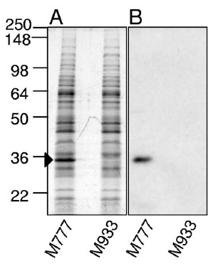
Although a lipL32 knockout mutant would help scientists figure out whether LipL32 plays an essential role in pathogenesis, targeted gene disruptions are extremely difficult with pathogenic Leptospira.  Fortunately, Ben Adler's group at Monash University obtained an insertion mutation in the lipL32 gene of L. interrogans by transposon mutagenesis.  This gave the Australians and their collaborators an opportunity to test the role of LipL32 in causing lethal infections in the hamster model of leptospirosis.  They first confirmed that the lipL32 mutant failed to express LipL32 by Western blotting the mutant with LipL32 antiserum.  I am showing the Coomassie-blue stained protein gel of the whole-cell lysate below so that you can appreciate the abundance of LipL32.  It is the most intensely stained band in the control L. interrogans strain, which has its lipL32 gene intact.

Whole-cell lysates of the L. interrogans lipL32 mutant (M933) and a control strain with the transposon in an intergenic region (M777) were run into SDS-acylamide gels and stained (panel A) or analyzed by Western blotting with LipL32 antiserum (panel B).  The M777 strain was demonstrated in an earlier study to be lethal for hamsters.  (Figure 1 from Murray et al., 2009.)

The survival curves show that the L. interrogans lipL32 mutant was just as lethal to hamsters as the parent L. interrogans with its lipL32 gene intact, irrespective of the infection route.  Hence, lipL32 is not necessary for lethal infections of hamsters, at least under the conditions used in this study.

Panel A: Groups of 8 hamsters were inoculated with 1,000 leptospires into the abdominal cavity.  Panel B:  Groups of 10 hamsters were inoculated with 106 leptospires dropped into the eye.  The slight difference in the survival curves was not statistically significant.  (Figure 5 from Murray et al., 2009)


Rats are the natural reservoir hosts of L. interrogans.  They can carry the spirochete for years in their kidney tubules without showing any signs of illness.  LipL32 could have a role in chronic infections.  The investigators therefore tested the ability of the lipL32 mutant to establish a chronic infection in laboratory rats.  They found that the lipL32 mutant (M933) was able to colonize the rat kidneys as well as the control M777 strain.  Kidneys from all 8 rats inoculated with the lipL32 mutant were culture positive.

At first glance it's surprising that lipL32 was not required for acute or chronic infection.  The authors pointed out that the function of LipL32 could be copied by other proteins found on the surface of Leptospira.  However, the study could have been strengthened by making two changes.  First, since the authors were trying to determine whether lipL32 was necessary for chronic infection, the rats should have been allowed to live for at least a few months before their kidneys were cultured.  Instead, the infection was allowed to proceed for only 15 days before the rats were sacrificed.  Second, they should have measured the bacterial load in the rat kidneys, either by plating serial dilutions of the kidney homogenates for colonies (although I don't know if this is feasible for Leptospira) or by quantitative PCR.  Clearly, more work needs to be done before anyone can conclude that LipL32 is not essential for chronic infection.


Featured paper

Murray G.L., Srikram, A., Hoke, D.E., Wunder Jr., E.A., Henry, R., Lo, M., Zhang, K., Sermswan, R.W., Ko, A.I., and Adler, B. (March 2009).  Major surface protein LipL32 is not required for either acute or chronic infection with Leptospira interrogansInfection and Immunity 77(3):952-958.  DOI: 10.1128/IAI.01370-08


Related posts

Saturday, July 24, 2010

Antigen presentation in the bloodstream: How invariant NKT cells are activated by Lyme disease spirochetes

The spirochete Borrelia burgdorferi is the tick-borne agent of Lyme disease, which affects the joints, nervous system, and heart.  After being deposited into the skin by an infected tick, the spirochete must enter the bloodstream so that it can circulate in the blood to gain access to its target organs.

The host doesn't sit idly as B. burgdorferi establishes an infection.  Invariant natural killer (iNKT) cells are one of the tools deployed by the immune system in its battle against the Lyme spirochetes.  Scientists know this because B. burgdorferi-infected mice lacking iNKT cells ended up with more spirochetes in their tissues and greater joint swelling than mice with a complete immune system.2

iNKT cells are an odd type of T cell.  Like other T cells, iNKT cells have a T cell receptor (TCR), yet they also express protein markers used to identify natural killer (NK) cells.  What makes the iNKT cell invariant is its TCR α chain, which comes only in the version dubbed Vα14 in mice and Vα24 in humans.  Even the β chain of the TCR of iNKT cells is restricted to three types in mice and just one in humans.  The lack of variation is unusual because the α and β TCR chains of conventional αβ T cells come in many forms in each individual, resulting in millions of varieties of TCRs.  This enables conventional αβ T cells to recognize a wide range of microbial peptide antigens when displayed by an MHC molecule on the surface of an antigen-presenting cell (see figure below).  In contrast, the TCRs of iNKT cells recognize a limited set of glycolipids displayed by the antigen-presenting cell's CD1d molecule, which structurally resembles MHC.  So far these glycolipids have been found only in Sphingomonas and B. burgdorferi.

Antigen recognition by T cells.  The "X" represents variable T cell receptor chains.
Figure 1 from ref. 3.
The structures of the B. burgdorferi glycolipids recognized by iNKT cells are shown below.  BbGL-IIc is recognized by mouse iNKT cells, and BbGL-IIf reacts with human iNKT cells.4
Structures of B. burgdorferi glycolipid antigens recognized by iNKT cells.  Figure 3d from ref. 3.

iNKT cells are activated when their TCR binds to BbGL-II complexed with CD1d.4  The activated iNKT cells secrete cytokines that elicit the appropriate immune response against the spirochetes.  How these cytokines promote killing of B. burgdorferi remains unknown.

To view the process of iNKT cell activation, scientists have recently obtained video footage of the early stages of the immune response to Borrelia burgdorferi circulating in the bloodstream of mice.1  The study by Lee et al., which appeared in the April issue of Nature Immunology, complements two earlier studies that revealed how the Lyme disease spirochete escapes from the bloodstream of mice to invade the surrounding tissues.5,6

The investigators employed fluorescence video microscopy to watch the immune cells in action following injection of an engineered B. burgdorferi strain expressing green fluorescent protein (GFP) into the bloodstream.  Although the spleen is better known for filtering bloodstream pathogens, the liver was selected for observation because iNKT cells make up 30% of the T cells in the liver.  In contrast, iNKT cells represent only 2.5% of T cells in the spleen.  Moreover, mice missing their spleen were able to limit B. burgdorferi infection as well as mice having a spleen, suggesting that the spleen is not critical in fighting bloodstream B. burgdorferi.

iNKT cells reside in the liver's sinusoids, which are the specialized capillaries that carry blood through the liver.  Similar to what other investigators have observed, the authors saw iNKT cells creeping along the inner surface of the liver sinusoids in healthy mice (see video below).

iNKT cells crawling within the liver sinusoids of a mouse genetically altered to express green fluorescent protein (GFP) in iNKT cells.  The iNKT cells glow bright green.  The elapsed time is shown at the top right.  Video 2 from ref. 1.

The investigators wanted to figure out which of the antigen-presenting cells found in the liver presented borrelial glycolipid to iNKT cells.  The answer?  After the spirochetes were injected into the bloodstream, they were quickly captured by Kupffer cells, the specialized blood-filtering macrophages that also reside in the liver sinusoids (see figure below).  Unlike iNKT cells, Kupffer cell remained stationary.

Capture of fluorescent B. burgdorferi (thin green bodies) by Kupffer cells (arrowhead).  Kupffer cells are stained red.  B. burgdorferi that avoided capture can be seen bound to the endothelium, trying to escape from the bloodstream into the liver tissue (arrow).  Figure 2e from ref. 1.

During the next several hours, the captured spirochetes were engulfed and broken up by the Kupffer cells so that BbGL-II could be loaded onto CD1d and displayed on the cell surface.  At 8 hours post injection, iNKT cells started to cluster and form stable contacts with Kupffer cells.  The iNKT cells were attracted to Kupffer cells churning out the chemokine CXCL9, a potent iNKT cell attractant.  The evidence for this was that injection of antibodies against the CXCL9 receptor, located on the iNKT cell surface, blocked clustering of iNKT cells.  Interaction of the Kupffer and iNKT cells was accompanied by increased blood and liver levels of the cytokine IFN-γ (interferon-gamma), a sign that the iNKT cells were being activated.

Left panel:  Liver 24 hours after injection of a GFP+ strain of B. burgdorferi into the bloodstream of a mouse.  Arrows indicate spirochetes (thin green bodies) that were not captured.  Kupffer cells are stained a red. The iNKT cells are the large bright green bodies.  The bright iNKT clusters overwhelm the faint red Kupffer cells, which are difficult to see.  Right panel:  To obtain a more convincing image showing contact between Kupffer cells and iNKT cells, a 3D reconstruction of the optical sections through the liver was performed.  Rotation of the image reveals interactions between Kupffer and iNKT cells.

Not all spirochetes were captured.  The investigators saw B. burgdorferi escaping from the sinusoids into the surrounding liver tissue even as other spirochetes were trapped by nearby Kupffer cells (see figures above).  Spirochetes circulating throughout the host probably escaped into other organs in the same manner.  Indeed, large amounts of  B. burgdorferi DNA were detected by PCR in several organs, including the liver, three days after the spirochetes were injected.  Although one doesn't usually think about the effects of Lyme disease on the liver, the authors pointed out that a mild hepatitis is common in Lyme disease patients.  In one prospective study, 40% of Lyme disease patients had at least one liver test abnormality.7

ResearchBlogging.orgBy now you may be wondering why the liver would devote such a high percentage of its T cells towards recognizing glycolipids that aren't found on most bacteria.  One answer is that microbes lacking the proper glycolipids may activate iNKT cells indirectly.3  For example, Salmonella typhimurium uses its LPS to coax antigen-presenting cells into making an endogenous glycolipid that gets presented to the iNKT cell by CD1d.8  It is also possible that glycolipids that are recognized by iNKT cells are present in other bacteria but are yet to be discovered.


Featured paper

1. Lee, W.Y., Moriarty, T.J., Wong, C.H.Y., Zhou, H., Strieter, R.M., van Rooijen, N., Chaconas, G., & Kubes, P. (2010). An intravascular immune response to Borrelia burgdorferi involves Kupffer cells and iNKT cells Nature Immunology, 11 (4), 295-302 DOI: 10.1038/ni.1855

Other references

2.  Tupin, E., Benhnia, M.R., Kinjo, Y., Patsey, R., Lena, C.J., Haller, M.C., Caimano, M.J., Imamura, M., Wong, C., Crotty, S., Radolf, J.D., Sellati, T.J., and Kronenberg, M. (2008).  NKT cells prevent chronic joint inflammation after infection with Borrelia burgdorferiProc. Natl. Acad. Sci. USA 105(50):19863-19868.  DOI: 10.1073/pnas.0810519105

3.  Tupin, E., Kinjo, Y., and Kronenberg, M. (2007).  The unique role of natural killer T cells in the response to microorganisms.  Nature Reviews Microbiology 5(6):405-417.  DOI: 10.1038/nrmicro1657

4.  Kinjo, J., Tupin, E., Wu, D., Fujio, M., Garcia-Navarro, R., Benhnia, M. R., Zajonc, D.M., Ben-Menachem, G., Ainge, G.D., Painter, G.F., Khurana, A., Hoebe, K., Behar, S.M., Beutler, B., Wilson, I.A., Tsuji, M., Sellati, T.J., Wong, C., and Kronenberg, M. (2006).  Nature Immunology 7(9):978-986.  DOI: 10.1038/ni1380

5.  Moriarty, T.J., Norman, M.U., Colarusso, P., Bankhead, T., Kubes, P., and Chaconas, G. (June 20, 2008).  Real-time high resolution 3D imaging of the Lyme disease spirochete adhering to and escaping from the vasculature of a living host.  PLoS Pathogens 4(6):e1000090.  DOI: 10.1371/journal.ppat.1000090

6. Norman, M.U., Moriarty, T.J., Dresser, A.R., Millen, B., Kubes, P., and Chaconas, G. (October 3, 2008). Molecular mechanisms involved in vascular interactions of the Lyme disease pathogen in a living host.  PLoS Pathogens 4(10):e1000169.  DOI: 10.1371/journal.ppat.1000169

7.  Horowitz, H.W.,  Dworkin, B., Forseter, G., Nadelman, R.B., Connolly, C., Luciano, B.B., Nowakowski, J., O'Brien, T.A., Calmann, M., Wormser, G.P. (June 1996).  Liver function in early Lyme disease.  Hepatology 23(6):1412-1417.  DOI: 10.1002/hep.510230617

8.  Mattner J., DeBord, K.L., Ismail, N., Goff, R.D., Cantu III, C., Zhou, D., Saint Mezard, P., Wang, V., Gao, Y., Yin, N, Hoebe, K., Schneewind, O., Walker, D., Beutler, B., Teyton, L, Savage, P.B., and Bendelac, A. (March 24, 2005).  Exogenous and endogenous glycolipid antigens activate NKT cells during microbial infections.  Nature 434(7032):525-529.  DOI: 10.1038/nature03408

Related posts